Regional Anatomy: Your Comprehensive Body Guide
Regional anatomy constitutes a pivotal approach in the broader field of anatomical study, emphasizing the organization of the human body into distinct parts or regions. Specifically, the University of Michigan Medical School integrates regional anatomy into its curriculum, providing medical students with a practical understanding of how structures relate within specific areas like the thorax or limbs. Furthermore, Gray's Anatomy, a historically significant and continually updated textbook, dedicates significant portions to explaining anatomical structures through a regional approach, facilitating a comprehensive understanding of their spatial relationships. In surgical planning, the utilization of imaging techniques, such as MRI scans, allows surgeons to visualize regional anatomy in detail, crucial for procedures requiring precise navigation through complex anatomical landscapes. Finally, clinical anatomists, such as Dr. John Smith, often specialize in regional anatomy, focusing their research and practice on specific body regions to advance diagnostic and therapeutic techniques.
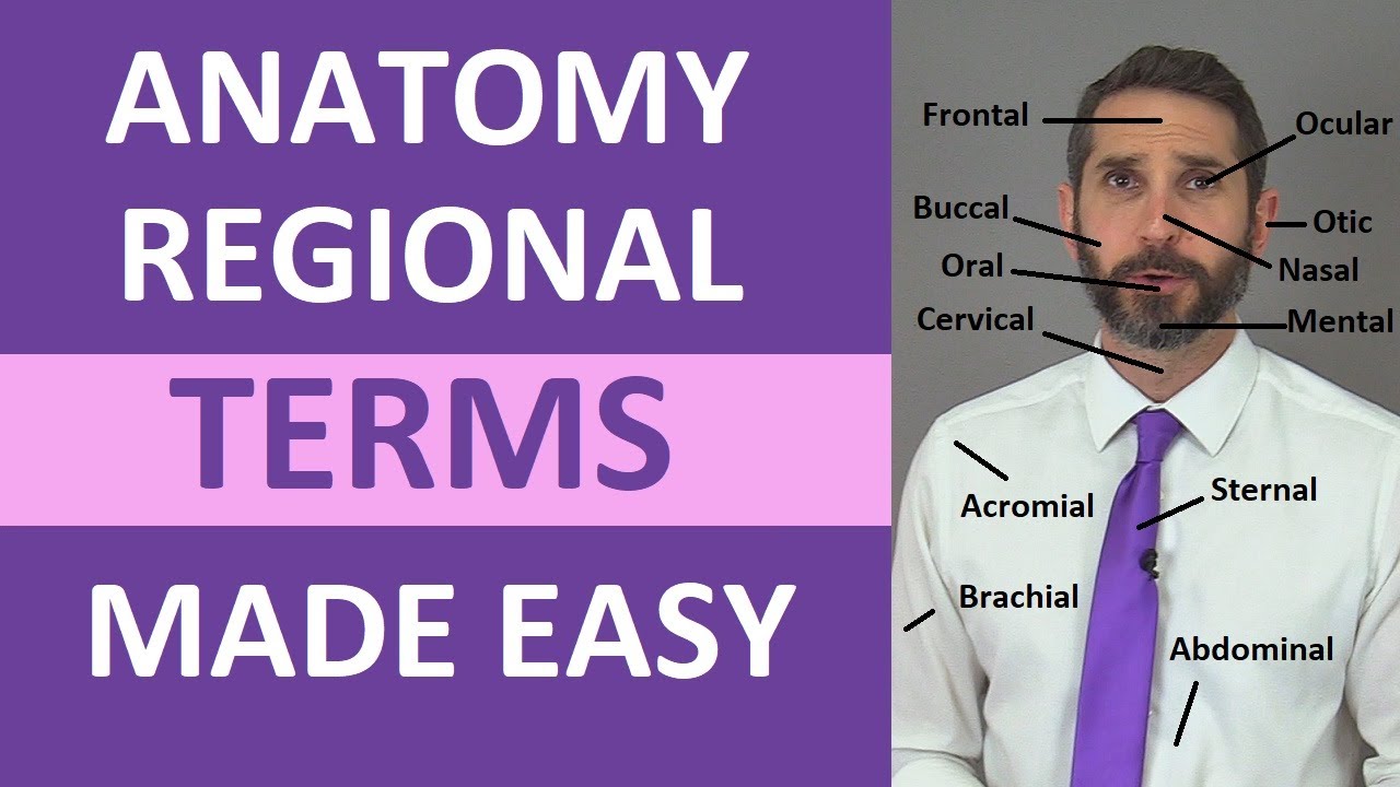
Image taken from the YouTube channel RegisteredNurseRN , from the video titled Regional Terms Anatomy - Body Parts Name | Nursing Medical Terminology Made Easy .
Unveiling the Wonders of Human Anatomy
Human anatomy, the scientific study of the body's structure, forms the bedrock of medical knowledge and biological understanding. It is far more than a mere catalog of parts; it is a dynamic field crucial for understanding health, disease, and the very essence of human existence. Its relevance extends far beyond textbooks and laboratories, influencing everything from surgical procedures to athletic performance.
The Profound Relevance of Anatomical Study
The significance of human anatomy resonates deeply across multiple disciplines.
For medical professionals, a thorough understanding of anatomy is indispensable. Surgeons rely on precise anatomical knowledge to navigate the body's intricate landscapes. Clinicians depend on it to accurately diagnose and treat ailments.
Even in seemingly unrelated fields like sports science, anatomical insights inform training regimens and injury prevention strategies.
A Comprehensive Overview
This editorial aims to provide a comprehensive overview of human anatomy, suitable for both students embarking on their anatomical journey and enthusiasts eager to deepen their appreciation of the human form. We will explore the historical milestones that have shaped our understanding of anatomy, paying homage to the pioneers whose dedication and discoveries have illuminated the inner workings of the body.
The editorial will delve into regional and systemic approaches to anatomical study. This will provide a framework for organizing complex information. It offers insights into anatomical terminology and essential concepts.
We will also discuss the tools and technologies that empower anatomical exploration, from traditional dissection to advanced imaging techniques. Finally, we will point to the resources that can serve as valuable guides for continued learning.
Goal: Empowering Understanding
Our primary goal is to empower readers with a foundational understanding of human anatomy. By demystifying its complexities, we hope to inspire a deeper appreciation for the intricate beauty and functional elegance of the human body. This editorial is designed to be a valuable resource for anyone seeking to unlock the secrets hidden beneath our skin.
Pioneers of Anatomy: Standing on the Shoulders of Giants
Following the initial understanding of the need to understand the human body, let us explore the historical figures who have been pivotal in shaping our understanding of anatomy. Their contributions have provided a foundation for modern medical science. We explore their groundbreaking work and enduring legacies.
The Ancient World: Foundations of Anatomical Thought
Anatomical inquiry dates back to ancient civilizations. These early explorations laid the groundwork for future discoveries.
While detailed records are scarce, figures like Hippocrates (c. 460 – c. 370 BC) made observations of the human body through dissection and clinical practice, emphasizing a holistic approach to medicine.
Aristotle (384–322 BC) further advanced anatomical knowledge through animal dissections, though his understanding of human anatomy was limited by the lack of human specimens. His work, however, established a framework for biological investigation that influenced scholars for centuries.
The Renaissance Revolution: Vesalius and the Dawn of Modern Anatomy
The Renaissance marked a turning point in anatomical study. This was characterized by a renewed interest in classical learning and direct observation.
Andreas Vesalius (1514–1564), a Flemish anatomist, revolutionized the field with his masterpiece, De humani corporis fabrica (1543).
Based on his dissections of human cadavers, Vesalius provided unprecedented accuracy and detail in anatomical illustrations, correcting many errors that had persisted for centuries from Galen’s teachings.
His meticulous work challenged established dogma and ushered in a new era of empirical observation and scientific rigor, earning him the title of the founder of modern anatomy.
The Artistic Anatomist: Leonardo da Vinci's Vision
Leonardo da Vinci (1452–1519), the quintessential Renaissance polymath, approached anatomy with an artist's eye and a scientist's curiosity.
Through meticulous dissections and detailed drawings, he sought to understand the human form in its entirety.
His anatomical studies, though largely unpublished during his lifetime, reveal a profound understanding of musculoskeletal anatomy, biomechanics, and the intricate relationships between different body systems.
Da Vinci's anatomical drawings are not only works of art, but also invaluable scientific documents that demonstrate his unparalleled observational skills and innovative approach to anatomical investigation.
Gray's Anatomy: A Timeless Classic
Published in 1858, Gray's Anatomy, authored by Henry Gray (1827–1861), became a cornerstone of medical education.
Its comprehensive descriptions and detailed illustrations of the human body have made it an indispensable resource for generations of medical students and healthcare professionals.
Despite being revised and updated numerous times, Gray's Anatomy remains a testament to Gray's meticulous scholarship and enduring legacy in the field of anatomy.
Modern Masters: Netter, Moore, and Agur
In the 20th and 21st centuries, anatomists like Frank Netter, Keith L. Moore, and Anne M.R. Agur have continued to advance the field through their contributions to anatomical illustration, textbook writing, and clinical anatomy.
Frank Netter (1906–1991), a physician and medical illustrator, created a vast collection of anatomical illustrations renowned for their clarity, accuracy, and artistic beauty.
His Atlas of Human Anatomy has become a standard reference work for medical students and clinicians worldwide.
Keith L. Moore (1925-2019) made significant contributions to clinical anatomy and embryology. His textbooks, such as Clinically Oriented Anatomy, emphasize the clinical relevance of anatomical knowledge.
Anne M.R. Agur is a contemporary anatomist known for her expertise in clinical anatomy and medical education. Her co-authorship of Grant's Atlas of Anatomy and contributions to other anatomical resources have made her a prominent figure in the field today.
Legacy and Inspiration
These pioneers of anatomy have left an indelible mark on the field. Their discoveries, illustrations, and teachings continue to shape our understanding of the human body and inspire future generations of anatomists and healthcare professionals.
Standing on the shoulders of these giants, we continue to explore the wonders of human anatomy, pushing the boundaries of knowledge and improving human health.
Regional Anatomy: Mapping the Human Body, Area by Area
Following the initial understanding of the need to understand the human body, let us explore the historical figures who have been pivotal in shaping our understanding of anatomy. Their contributions have provided a foundation for modern medical science. We explore their groundbreaking work and lasting legacy, now let's transition to a crucial aspect of anatomical study: regional anatomy. This approach focuses on examining the human body by dividing it into distinct areas. This method allows for a comprehensive understanding of the relationships between various structures within a specific region.
Exploring Anatomical Regions
Regional anatomy involves studying the body by area, allowing for a deep dive into the interconnections of different systems within each region. This method is particularly useful in clinical settings where a specific area of the body is of concern. Let us examine the major regions and their key anatomical features.
Head and Neck: The Seat of Senses and Control
The head and neck region contains some of the most complex anatomy in the human body. It houses the brain, sensory organs, and the upper portions of the respiratory and digestive tracts.
Understanding the intricate network of nerves, blood vessels, and muscles in this area is crucial for diagnosing and treating neurological and sensory disorders. The density of structures in this region necessitates a thorough understanding of spatial relationships.
Thorax (Chest): Protecting Life-Sustaining Organs
The thoracic region, or chest, is primarily responsible for housing and protecting vital organs such as the heart and lungs. The bony rib cage, intercostal muscles, and mediastinum are key components of this region.
Knowledge of the anatomical relationships within the thorax is essential for cardiologists, pulmonologists, and surgeons performing procedures in this area.
Abdomen: The Center of Digestion and Metabolism
The abdominal region contains the majority of the digestive organs, including the stomach, intestines, liver, pancreas, and spleen. This area is responsible for processing nutrients, eliminating waste, and regulating various metabolic processes.
Understanding the arrangement and function of these organs is crucial for diagnosing and treating gastrointestinal disorders. The complexity of this area requires a systematic approach to studying its anatomy.
Pelvis: Housing Reproductive and Urinary Systems
The pelvic region houses the reproductive and urinary systems, including the bladder, rectum, and internal reproductive organs. This area is essential for reproduction, urinary excretion, and maintaining continence.
Knowledge of pelvic anatomy is vital for gynecologists, urologists, and colorectal surgeons. The anatomical differences between male and female pelvises also necessitate careful study.
Upper Limb: Enabling Dexterity and Manipulation
The upper limb, consisting of the shoulder, arm, forearm, and hand, is specialized for dexterity and manipulation. The intricate arrangement of bones, muscles, nerves, and blood vessels allows for a wide range of movements and fine motor skills.
Understanding the biomechanics of the upper limb is crucial for physical therapists, orthopedic surgeons, and occupational therapists.
Lower Limb: Supporting Weight and Facilitating Movement
The lower limb, comprising the hip, thigh, leg, and foot, is responsible for supporting weight and facilitating movement. The strong bones, powerful muscles, and complex joints of the lower limb enable walking, running, and jumping.
Knowledge of lower limb anatomy is essential for podiatrists, orthopedic surgeons, and sports medicine physicians. The weight-bearing function of this limb makes it particularly susceptible to injury and degeneration.
Back (Spine): Providing Support and Protection
The back, or spine, provides structural support for the body and protects the spinal cord. The vertebral column, intervertebral discs, and associated muscles allow for flexibility and stability.
Understanding the anatomy of the back is crucial for chiropractors, neurosurgeons, and pain management specialists. The intricate network of nerves and muscles in the back makes it a common site for pain and injury.
Advantages of Studying Regional Anatomy
Studying anatomy from a regional perspective offers several advantages. It allows for a comprehensive understanding of the relationships between different structures within a specific area.
This approach is particularly useful in clinical practice, where problems often manifest in a localized region. Furthermore, regional anatomy promotes a holistic understanding of the body. It highlights the interconnectedness of different systems and their functions.
By studying anatomy regionally, students and professionals can develop a deeper appreciation for the complexity and efficiency of the human body. This perspective allows for more effective diagnosis, treatment, and management of various medical conditions.
Systemic Anatomy: Understanding Functional Networks
Following our exploration of regional anatomy, which segments the body into discrete areas, we turn our attention to systemic anatomy. This approach provides a holistic view, examining the body through the lens of its integrated organ systems. Rather than focusing on location, systemic anatomy emphasizes the functional unity of structures that work together to perform specific vital processes.
This perspective is crucial for comprehending the complex interplay that sustains life. It allows us to appreciate how seemingly disparate parts of the body are interconnected and interdependent.
The Major Systems: A Functional Overview
Systemic anatomy divides the body into eleven principal organ systems. Each system comprises a collection of organs and tissues that collaborate to execute essential functions.
Let's delve into these systems, exploring their individual roles and their contributions to the body's overall well-being.
Musculoskeletal System: Framework for Movement
The musculoskeletal system provides the body's structural support and enables movement. It is an integrated network of bones, muscles, joints, tendons, and ligaments. Bones provide a rigid framework, while muscles generate the force needed for motion. Joints are the articulations where bones meet, allowing for a range of movements, while ligaments connect bone to bone.
Tendons attach muscles to bones. This system not only facilitates movement but also protects vital organs and maintains posture.
Nervous System: Command and Control
The nervous system acts as the body's control center, coordinating and regulating bodily functions. It consists of the brain, spinal cord, nerves, and sensory receptors. The brain serves as the central processing unit, while the spinal cord acts as the main communication pathway. Nerves transmit signals throughout the body, allowing for rapid communication between different regions.
This system is responsible for sensation, perception, thought, and voluntary and involuntary actions.
Cardiovascular System: Transport and Delivery
The cardiovascular system is responsible for transporting blood, oxygen, nutrients, and hormones throughout the body. It comprises the heart, blood vessels (arteries, veins, and capillaries), and blood. The heart pumps blood through the circulatory system, while arteries carry oxygenated blood away from the heart.
Veins return deoxygenated blood to the heart. This system ensures that all tissues receive the necessary resources for survival and removes metabolic waste products.
Lymphatic System: Immunity and Fluid Balance
The lymphatic system plays a vital role in immunity and fluid balance. It consists of lymph nodes, lymphatic vessels, and lymphoid organs such as the spleen and thymus. Lymph nodes filter lymph fluid, removing pathogens and cellular debris. The lymphatic system also returns excess fluid from tissues back into the bloodstream.
This system is essential for maintaining fluid homeostasis and defending the body against infection.
Respiratory System: Gas Exchange
The respiratory system facilitates gas exchange, bringing oxygen into the body and expelling carbon dioxide. It includes the lungs, trachea, bronchi, and diaphragm. The lungs are the primary site of gas exchange, where oxygen is absorbed into the blood and carbon dioxide is released. The diaphragm, a major muscle of respiration, contracts and relaxes to facilitate breathing.
This system is crucial for sustaining cellular respiration and energy production.
Digestive System: Nutrient Processing
The digestive system breaks down food into smaller molecules that can be absorbed into the bloodstream. It consists of the mouth, esophagus, stomach, small intestine, large intestine, liver, pancreas, and gallbladder. Each organ plays a specific role in the digestive process, from mechanical breakdown to enzymatic digestion.
The small intestine is the primary site of nutrient absorption, while the large intestine absorbs water and electrolytes. This system provides the body with the nutrients it needs for growth, repair, and energy.
Urinary System: Waste Elimination
The urinary system filters waste products from the blood and eliminates them from the body as urine. It comprises the kidneys, ureters, bladder, and urethra. The kidneys are the primary filtering organs, removing toxins, excess salts, and other waste products from the bloodstream. The ureters transport urine from the kidneys to the bladder.
The bladder stores urine until it is eliminated through the urethra. This system maintains fluid and electrolyte balance and removes harmful substances from the body.
Reproductive System: Procreation
The reproductive system enables procreation and the continuation of the species. It differs between males and females, with specialized organs and structures. The male reproductive system produces sperm, while the female reproductive system produces eggs. Fertilization occurs when sperm and egg unite, leading to the development of a new organism.
This system is essential for the survival of the species and the transmission of genetic information.
Endocrine System: Hormone Regulation
The endocrine system regulates bodily functions through the secretion of hormones. It consists of various glands, including the pituitary gland, thyroid gland, adrenal glands, pancreas, and ovaries or testes. Hormones act as chemical messengers, traveling through the bloodstream to target cells and tissues.
This system controls growth, metabolism, reproduction, and other essential functions.
Integumentary System: Protection and Sensation
The integumentary system, comprising the skin, hair, and nails, serves as the body's outer protective layer. The skin acts as a barrier against infection, injury, and dehydration. It also regulates body temperature and provides sensory information. Hair and nails offer additional protection and support.
This system is vital for maintaining homeostasis and interacting with the external environment.
Advantages of a Systemic Approach
Understanding the human body through a systemic approach offers several key advantages.
It promotes a deeper appreciation of how different organs and tissues work together to maintain overall health and function. This approach is essential for diagnosing and treating diseases, as it allows healthcare professionals to understand how a problem in one system can affect other parts of the body.
By studying the body's systems, we can gain a more complete and integrated understanding of human biology.
Tools of the Trade: Essential Technologies for Anatomical Exploration
The study of human anatomy has been profoundly shaped by technological advancements, transforming our understanding of the human body from macroscopic observations to intricate microscopic details. From the age-old practice of dissection to the sophisticated realm of medical imaging, a diverse array of tools and techniques empower anatomists, researchers, and students to explore the complexities of the human form. Let us delve into these essential instruments that unveil the secrets within.
The Art and Science of Dissection
Dissection, the meticulous process of carefully separating and examining tissues, stands as the cornerstone of anatomical study. It is the quintessential hands-on method, providing an unparalleled opportunity to directly observe the spatial relationships and structural characteristics of organs, vessels, and nerves.
While modern technology has undoubtedly enhanced anatomical education, the irreplaceable value of dissection lies in the tactile and visual experience it offers. This direct engagement fosters a deeper understanding that cannot be replicated by virtual simulations alone.
Visualizing the Microscopic World: Microscopy
Microscopy opens the door to a hidden world, revealing the intricate architecture of tissues and cells. This is at a level beyond the capabilities of the naked eye. From basic light microscopy to advanced electron microscopy, these techniques allow us to visualize cellular structures, identify tissue types, and understand the underlying mechanisms of disease.
Histology, the study of tissues, relies heavily on microscopy for the identification and classification of cellular components. This method offers invaluable insights into both normal anatomy and pathological changes.
Imaging Technologies: A Window into the Living Body
The advent of medical imaging has revolutionized anatomical study, providing non-invasive methods for visualizing the internal structures of the living body. Each imaging modality offers a unique perspective, enabling clinicians and researchers to diagnose, monitor, and treat a wide range of conditions.
X-rays (Radiography): Illuminating the Skeletal Framework
Radiography, utilizing X-rays to penetrate tissues, is particularly effective for visualizing bones, detecting fractures, and identifying abnormalities in skeletal structures. While offering limited detail for soft tissues, X-rays remain an invaluable tool for initial assessments and quick diagnoses.
Computed Tomography (CT Scans): Constructing Cross-Sectional Images
Computed tomography (CT) scans employ X-rays and computer processing to generate detailed cross-sectional images of the body. This technique provides a more comprehensive view than traditional radiography, allowing visualization of soft tissues, organs, and blood vessels. CT scans are particularly useful for diagnosing internal injuries, detecting tumors, and guiding surgical procedures.
Magnetic Resonance Imaging (MRI): Soft Tissue Visualization with Precision
Magnetic resonance imaging (MRI) uses powerful magnets and radio waves to create detailed images of soft tissues. MRI excels in visualizing the brain, spinal cord, muscles, ligaments, and other soft tissue structures, providing superior contrast and resolution compared to CT scans. This non-invasive imaging modality is essential for diagnosing neurological disorders, musculoskeletal injuries, and a variety of other conditions.
Ultrasound: Real-Time Imaging through Sound Waves
Ultrasound utilizes high-frequency sound waves to create real-time images of internal structures. This portable and relatively inexpensive imaging technique is commonly used for prenatal imaging, assessing blood flow, and guiding needle biopsies. Ultrasound's real-time capability makes it particularly useful for visualizing moving structures, such as the heart and blood vessels.
Anatomical Models: Physical Representations for Enhanced Understanding
Anatomical models provide tangible representations of the human body, enhancing spatial understanding and facilitating learning. These models range from simple plastic skeletons to highly detailed replicas of individual organs and systems.
The advantage of anatomical models lies in their ability to be manipulated and examined from multiple angles, allowing students to grasp complex anatomical relationships more easily. These models serve as invaluable tools for both introductory and advanced anatomical study.
Anatomy Software: Interactive Learning in the Digital Age
Anatomy software provides interactive platforms for exploring the human body in a virtual environment. These programs often feature 3D models, interactive dissections, and detailed anatomical annotations.
Anatomy software offers several advantages, including the ability to rotate and manipulate structures, peel away layers to reveal deeper anatomy, and test knowledge through quizzes and simulations. These digital tools are revolutionizing anatomical education, making learning more engaging and accessible.
The tools and technologies used in anatomical exploration are constantly evolving, driven by scientific advancements and technological innovation. From traditional dissection to cutting-edge imaging modalities and interactive software, these resources empower us to delve deeper into the mysteries of the human body, ultimately improving healthcare, advancing scientific knowledge, and fostering a greater appreciation for the intricate design of human life.
Navigating the Anatomical Landscape: Key Concepts and Terminology
The study of human anatomy has been profoundly shaped by technological advancements, transforming our understanding of the human body from macroscopic observations to intricate microscopic details. From the age-old practice of dissection to the sophisticated realm of medical imaging, these tools empower us to explore the complex architecture of the human form. However, regardless of the tools employed, a firm grasp of the fundamental concepts and precise terminology is paramount for effective anatomical study and communication.
The Importance of Anatomical Language
Anatomical terminology is not merely a collection of labels; it is a standardized, universally accepted language. This language allows healthcare professionals, researchers, and students across the globe to communicate accurately and unambiguously about the structure and organization of the human body. Without this shared vocabulary, misinterpretations and errors could have severe consequences in clinical settings, research endeavors, and educational programs.
Foundational Concepts in Anatomical Description
Several core concepts underpin all anatomical descriptions. These concepts provide a framework for understanding spatial relationships and orientations within the body.
Anatomical Position: The Starting Point
All anatomical descriptions are based on the anatomical position: a standardized reference point. In this position, the body is erect, feet flat on the floor, arms at the sides, and palms facing forward. This universally accepted convention ensures consistency and clarity in anatomical discourse.
Planes of Section: Slicing Through Complexity
To view internal structures, anatomists use planes of section: imaginary flat surfaces that pass through the body. The three primary planes are:
-
Sagittal: Divides the body into right and left parts. A midsagittal plane divides the body into equal halves.
-
Coronal (Frontal): Divides the body into anterior (front) and posterior (back) parts.
-
Transverse (Horizontal or Axial): Divides the body into superior (upper) and inferior (lower) parts.
Understanding these planes is crucial for interpreting medical images and visualizing anatomical structures in three dimensions.
Directional Terms: Describing Relationships
Directional terms are used to describe the relative position of one structure to another. Key directional terms include:
-
Superior (Cranial): Toward the head or upper part of a structure.
-
Inferior (Caudal): Away from the head or toward the lower part of a structure.
-
Anterior (Ventral): Toward the front of the body.
-
Posterior (Dorsal): Toward the back of the body.
-
Medial: Toward the midline of the body.
-
Lateral: Away from the midline of the body.
-
Proximal: Closer to the point of attachment or origin.
-
Distal: Farther from the point of attachment or origin.
-
Superficial: Closer to the surface of the body.
-
Deep: Away from the surface of the body.
Mastering these terms is essential for accurately describing the location of anatomical structures.
Body Cavities: Housing the Organs
The body contains several cavities that protect and support internal organs. The two main cavities are:
-
Dorsal Cavity: Located on the posterior aspect of the body, houses the cranial cavity (containing the brain) and the vertebral canal (containing the spinal cord).
-
Ventral Cavity: Located on the anterior aspect of the body, it is subdivided into the thoracic cavity (containing the heart and lungs) and the abdominopelvic cavity. The abdominopelvic cavity is further divided into the abdominal cavity (containing digestive organs, kidneys) and the pelvic cavity (containing reproductive organs, bladder, and rectum).
Specialized Anatomical Disciplines
Anatomy encompasses several specialized sub-disciplines, each focusing on a particular aspect of the body's structure:
Osteology: The Framework of the Body
Osteology is the study of bones. This discipline involves examining the structure, function, and development of the skeletal system. Key areas of study include bone morphology, bone growth, and the classification of different bone types.
Myology: The Machinery of Movement
Myology is the study of muscles. It focuses on the structure, function, and classification of different types of muscles (skeletal, smooth, and cardiac). Understanding myology is crucial for comprehending how the body moves and maintains posture.
Arthrology: Where Bones Meet
Arthrology is the study of joints. It explores the structure, function, and classification of different types of joints, including fibrous, cartilaginous, and synovial joints. Understanding arthrology is essential for comprehending how bones articulate and allow for movement.
Neurology: The Body's Control Center
Neurology is the study of the nervous system. This intricate system controls and coordinates bodily functions, and neurology delves into the structure and function of the brain, spinal cord, and peripheral nerves.
Angiology: The Circulatory Network
Angiology is the study of blood vessels and lymphatic vessels. It focuses on the structure and function of arteries, veins, capillaries, and lymphatic vessels, which are essential for transporting blood, nutrients, and waste throughout the body.
The Imperative of Precise Terminology
In conclusion, a thorough understanding of anatomical concepts and terminology is indispensable for anyone studying or working in the health sciences. The ability to communicate clearly and accurately about anatomical structures is crucial for effective clinical practice, research, and education. Embracing this specialized language unlocks deeper insights into the complexities of the human body and its remarkable functions.
Organizations and Resources: Your Guide to Anatomical Information
Navigating the Anatomical Landscape: Key Concepts and Terminology The study of human anatomy has been profoundly shaped by technological advancements, transforming our understanding of the human body from macroscopic observations to intricate microscopic details. From the age-old practice of dissection to the sophisticated realm of medical imaging, the resources available to students and professionals are extensive and ever-evolving. This section serves as a guide to some of the most valuable organizations, publications, and digital platforms that can significantly enhance anatomical knowledge and proficiency.
Key Organizations and Institutions
Several organizations and institutions stand as pillars of anatomical research, education, and dissemination. These entities provide invaluable resources, from comprehensive databases to hands-on training programs, catering to diverse needs within the anatomical sciences.
National Library of Medicine (NLM) and the Visible Human Project
The National Library of Medicine (NLM), a branch of the National Institutes of Health, is a cornerstone for medical information.
Its Visible Human Project is particularly noteworthy, offering detailed, digitized anatomical data derived from complete male and female cadavers.
This project serves as a crucial reference for researchers and educators, providing a virtual dissection resource that complements traditional methods.
Medical Schools & Universities: Departments of Anatomy
Departments of Anatomy at medical schools and universities are central to anatomical education and research.
These departments not only provide formal training to medical, dental, and allied health students, but they also engage in cutting-edge research.
Many departments offer open educational resources, including online lectures, interactive models, and anatomical atlases, accessible to a broader audience.
Anatomical Societies
Societies such as the American Association for Anatomy (AAA) and international counterparts provide a platform for collaboration, knowledge exchange, and professional development within the anatomical community.
These societies hold conferences, publish journals, and offer educational resources to promote anatomical research and education.
Essential Textbooks and Publishers
The foundation of anatomical knowledge often rests on meticulously crafted textbooks that present complex information in a structured and accessible manner. The following publishers are recognized for their contributions to anatomical literature:
Elsevier
Elsevier publishes some of the most respected anatomy textbooks, including Gray's Anatomy for Students and Netter's Atlas of Human Anatomy.
These resources are known for their comprehensive coverage and high-quality illustrations, making them essential for students and practitioners alike.
Wolters Kluwer
Wolters Kluwer is another prominent publisher of medical textbooks, featuring Moore's Clinically Oriented Anatomy and Rohen's Color Atlas of Anatomy.
These books emphasize the clinical relevance of anatomical knowledge, bridging the gap between basic science and clinical practice.
Online Learning Platforms and Digital Resources
The digital age has transformed anatomical education, providing interactive and engaging learning experiences that complement traditional methods.
3D Anatomy Software
Software platforms like Visible Body and Complete Anatomy offer interactive 3D models of the human body.
These tools allow users to explore anatomical structures from various angles, dissect virtual specimens, and test their knowledge through quizzes and assessments.
Anatomical Websites and Online Atlases
Websites such as Anatomography and online anatomical atlases provide free access to detailed anatomical images and information.
These resources can be valuable supplements to textbooks and lectures, offering a visual and interactive way to learn anatomy.
YouTube Channels and Educational Videos
Platforms like YouTube host numerous channels dedicated to anatomical education, featuring lectures, dissections, and tutorials.
These videos can provide alternative explanations of complex concepts and offer a visual learning experience that appeals to a wide range of learners.
The Role of Libraries and Archives
Libraries and archives play a crucial role in preserving and providing access to anatomical knowledge.
Medical libraries, in particular, offer extensive collections of anatomical textbooks, journals, and historical documents, serving as valuable resources for researchers and students.
Digital archives are increasingly important, preserving and making accessible historical anatomical illustrations and texts.
Ethical Considerations in Utilizing Resources
When utilizing anatomical resources, it is essential to consider ethical implications.
Access to cadaveric materials, for example, should be governed by ethical guidelines that respect the dignity of the deceased.
Similarly, the use of digital anatomical models should be accompanied by an understanding of their limitations and potential for misinterpretation.
Anatomy in Context: Interdisciplinary Connections
[Organizations and Resources: Your Guide to Anatomical Information Navigating the Anatomical Landscape: Key Concepts and Terminology The study of human anatomy has been profoundly shaped by technological advancements, transforming our understanding of the human body from macroscopic observations to intricate microscopic details. From the age-old pra...]
The true power of anatomical knowledge lies not in its isolated study, but in its synergistic integration with other scientific and medical disciplines. Anatomy forms the bedrock upon which our understanding of the human body, in both health and disease, is constructed. Its connections are far-reaching and essential to numerous fields.
This section elucidates these vital interdisciplinary relationships.
Anatomy and Histology: The Microscopic World
Histology, the study of tissues at the microscopic level, provides essential context to anatomical structures. While anatomy reveals the form and arrangement of organs, histology unveils their cellular composition and organization.
Understanding the specific types of cells that comprise a tissue is crucial for interpreting its function within an organ and its role in the overall body system.
For example, the anatomical structure of the small intestine, with its villi and microvilli, is only fully appreciated when combined with the histological knowledge of the absorptive enterocytes that line its surface.
Anatomy and Embryology: Unraveling Development
Embryology, the study of prenatal development, offers profound insights into the origins and formation of anatomical structures.
Understanding the embryological development of the heart, for instance, is essential for comprehending congenital heart defects.
Deviations during the intricate processes of embryogenesis can lead to a wide range of anatomical variations and malformations, underscoring the critical link between developmental biology and anatomical understanding.
Anatomy and Physiology: Form Dictates Function
Physiology, the study of bodily functions, is intimately linked to anatomy. Structure dictates function, and a comprehensive understanding of anatomy is essential for interpreting physiological processes.
The intricate anatomical design of the lungs, with their vast network of alveoli, directly facilitates the efficient exchange of gases during respiration.
Conversely, understanding physiological requirements can inform our understanding of why certain anatomical structures have evolved to their specific form.
Clinical Anatomy: Bridging the Gap
Clinical anatomy directly applies anatomical knowledge to the practice of medicine. Clinicians rely on a detailed understanding of anatomy to diagnose and treat diseases, perform surgical procedures, and interpret medical imaging.
Surgical approaches are guided by precise knowledge of anatomical landmarks, and the effectiveness of many medical interventions depends on a thorough understanding of anatomical relationships.
Clinical anatomy represents the practical culmination of anatomical study.
Anatomy and Radiology: Visualizing the Interior
Radiology, the use of medical imaging techniques such as X-rays, CT scans, MRI, and ultrasound, relies heavily on anatomical knowledge. Radiologists must be able to identify and interpret anatomical structures on these images to diagnose pathology.
A deep understanding of normal anatomical variations is crucial for distinguishing normal from abnormal findings.
Radiological anatomy has become an indispensable tool in modern medical practice.
Anatomy and Surgery: The Art of Precision
Surgery demands an exceptional level of anatomical knowledge and precision. Surgeons must possess a thorough understanding of anatomical structures, their relationships, and potential variations.
Surgical procedures require careful planning and execution, guided by anatomical landmarks and principles.
The success of surgical interventions is directly linked to the surgeon's mastery of anatomy.
Anatomy and Physical Therapy: Restoring Movement
Physical therapists utilize anatomical knowledge to assess and treat musculoskeletal conditions. They must understand the anatomical relationships between muscles, bones, joints, and nerves to design effective rehabilitation programs.
Knowledge of biomechanics, which is rooted in anatomy, is essential for understanding human movement and developing strategies to restore function.
Physical therapy relies heavily on a deep understanding of functional anatomy.
The Foundation of Medical Science
Anatomy serves as a cornerstone for a vast array of medical and scientific disciplines. Its importance cannot be overstated.
From understanding the intricacies of cellular function to performing complex surgical procedures, anatomical knowledge is fundamental to advancing our understanding of the human body and improving human health. Anatomy is not merely a subject of study, but a lens through which we view the very essence of human life.
Video: Regional Anatomy: Your Comprehensive Body Guide

FAQs: Regional Anatomy: Your Comprehensive Body Guide
What exactly is "regional anatomy" as covered in this guide?
Regional anatomy focuses on studying the body by specific areas, such as the head and neck or the upper limb. Unlike systemic anatomy, it emphasizes the relationships between all the structures within a defined region, including muscles, nerves, vessels, and bones. This guide provides a comprehensive understanding of these regions.
Who would benefit most from using this body guide?
Students in medical, nursing, and allied health programs will find this guide invaluable for learning regional anatomy. Practicing clinicians can also use it as a quick reference. Anyone curious about the structure and function of the human body will gain knowledge from this comprehensive resource.
How does this guide differ from other anatomy resources?
This guide focuses on a regional approach, prioritizing understanding the complex relationships within specific body regions. It provides detailed descriptions and clear visuals, unlike some resources that only focus on systemic anatomy (organs). The guide emphasizes the practical application of regional anatomy knowledge.
What if I’m unfamiliar with basic anatomical terminology?
While the guide aims to be comprehensive, a basic understanding of anatomical terms will be helpful. The book includes a glossary of key terms. If you are completely new to anatomy, consider reviewing an introductory anatomy resource alongside this guide for a smoother learning experience.
So, there you have it! Hopefully, this overview of regional anatomy has given you a better appreciation for how intricately connected your body really is. Keep exploring, keep learning, and remember that understanding regional anatomy is like having a map to your own incredible inner landscape.
