Epidural Needle Size in Inches: Your Guide
The gauge of an epidural needle, typically ranging between 16 and 18, represents a critical factor in determining the ease of insertion and minimizing the risk of complications during epidural anesthesia. Obstetricians, who administer epidurals to alleviate labor pain, rely on precise measurements like epidural needle size in inches to ensure optimal patient comfort and safety. The Tuohy needle, a specific type of epidural needle characterized by its curved tip, facilitates controlled advancement through the epidural space. Understanding the millimeter-to-inch conversion for these needles is essential, as variations in size, even fractions of an inch, can significantly impact the success and safety of the procedure, particularly when considering factors such as the patient's Body Mass Index (BMI).
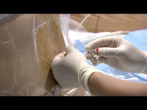
Image taken from the YouTube channel BabyCenter , from the video titled Epidural injection procedure .
Understanding the Critical Role of Epidural Needle Size in Pain Management
Epidural anesthesia and analgesia stand as cornerstones in modern pain management, offering targeted relief through the injection of anesthetic agents into the epidural space. This space, located just outside the dura mater (the membrane surrounding the spinal cord), becomes the conduit for medication to block nerve signals, alleviating pain in specific regions of the body.
The primary purpose of an epidural is to provide analgesia (pain relief) or anesthesia (loss of sensation) without inducing complete unconsciousness. This targeted approach allows patients to remain alert and participate in their care, a significant advantage in many clinical scenarios.
Epidurals: A Versatile Tool in Pain Management
Epidural techniques play a vital role across a broad spectrum of medical needs:
-
Labor and Delivery: Epidurals are frequently used to manage labor pain, providing significant relief for expectant mothers.
-
Post-operative Pain: Following major surgeries, epidurals can deliver continuous pain relief, facilitating recovery and reducing reliance on opioid medications.
-
Chronic Pain Management: In cases of persistent pain conditions, such as back pain or cancer pain, epidural injections may offer long-term relief and improved quality of life.
-
Specific Surgical Procedures: Epidurals are utilized as the primary anesthetic or as an adjunct to general anesthesia for surgeries involving the lower body and extremities.
The Paramount Importance of Correct Needle Size
While the epidural procedure itself is well-established, the seemingly simple act of selecting the appropriate needle size carries profound implications for both the effectiveness of the pain relief and the safety of the patient. The needle serves as the crucial bridge to deliver medication to the epidural space; therefore, choosing the correct size is of utmost importance.
Impact on Effectiveness
If a needle is too short, it may not reach the epidural space reliably, leading to inadequate pain relief and the need for repeated attempts. This can increase patient discomfort and prolong the procedure.
Conversely, a needle that is too long may increase the risk of unintended dural puncture, leading to a spinal headache, a potentially debilitating complication.
Safety and Minimizing Complications
The gauge (diameter) of the needle also plays a critical role in patient safety. Larger gauge needles increase the risk of bleeding and nerve damage. Smaller needles, while potentially less traumatic, may be more prone to deflection and can be more difficult to advance through tougher tissues.
Therefore, a nuanced understanding of needle dimensions and their correlation to patient anatomy and procedure type is essential for anesthesiologists to optimize outcomes and minimize potential complications. This requires a careful evaluation of each patient and a thorough understanding of the equipment being used.
Decoding Epidural Needle Size: Length and Diameter
Navigating the intricacies of epidural procedures demands a precise understanding of the tools at hand. Among these, the epidural needle itself is paramount, and its dimensions—length and diameter—are critical determinants of success and safety.
These seemingly simple measurements dictate the needle's reach, maneuverability, and potential impact on surrounding tissues. A thorough grasp of these parameters is thus essential for every practitioner.
Understanding Length Measurements
The length of an epidural needle directly influences its ability to reach the epidural space, a critical factor considering variations in patient anatomy and body habitus. Standard lengths are typically expressed in both inches and millimeters to facilitate international collaboration and consistent documentation.
Standard Lengths in Inches
Epidural needles are available in a range of lengths, typically varying from 2.5 inches to 4 inches. The selection depends primarily on the patient's size, the depth of the epidural space, and the chosen approach (midline vs. paramedian).
A shorter needle might suffice for a thin patient with a readily accessible epidural space, while a longer needle becomes necessary for individuals with greater tissue depth or when navigating through a more oblique angle.
Converting to Millimeters for Precision
For increased precision and consistency, especially in research and international settings, these lengths are often expressed in millimeters. A 2.5-inch needle corresponds to approximately 63.5 mm, while a 4-inch needle measures about 101.6 mm.
This conversion allows for finer adjustments and a more standardized approach to needle selection, minimizing potential errors arising from unit conversions.
Deciphering Diameter: The Gauge System
The diameter of an epidural needle, crucial for its ease of insertion and potential for complications, is measured using the gauge (G) system. Understanding this system is vital, as it dictates the needle's outer diameter and its impact on tissue displacement.
The Gauge System Explained
The gauge system operates on an inverse scale: the higher the gauge number, the smaller the needle's outer diameter. This counterintuitive relationship can be initially confusing, but it is essential to master.
For instance, a 18G needle has a smaller outer diameter than a 16G needle. It is important to remember this inverse relationship.
Gauge and Outer Diameter: A Critical Relationship
While the gauge number provides a relative indication of size, the actual outer diameter in millimeters offers a more precise understanding. An 18G epidural needle typically has an outer diameter of approximately 1.27 mm, while a 16G needle measures around 1.65 mm.
This difference, though seemingly small, can significantly impact the force required for insertion and the potential for tissue trauma. Choosing the appropriate gauge involves balancing the need for adequate needle stiffness with the desire to minimize tissue disruption.
Exploring Different Types of Epidural Needles: Tuohy vs. Crawford
Navigating the intricacies of epidural procedures demands a precise understanding of the tools at hand. Among these, the epidural needle itself is paramount, and its dimensions—length and diameter—are critical determinants of success and safety.
These seemingly simple measurements dictate the needle's maneuverability, its ability to access the epidural space, and, ultimately, the risk of complications.
While needle size is a key factor, the design of the needle tip plays an equally vital role. Two prominent designs dominate the field: the Tuohy needle and the Crawford needle.
Each offers distinct advantages and disadvantages, making them suitable for specific clinical scenarios.
The Tuohy Needle: A Curved Advantage
The Tuohy needle, characterized by its curved or slightly beveled tip, is arguably the more widely recognized and utilized of the two.
Its curved design offers several key advantages, primarily related to its ability to deflect rather than cut through tissues.
This characteristic is particularly beneficial when advancing the needle through the ligaments and tissues of the back towards the epidural space.
Deflection and Directional Control
The curved tip of the Tuohy needle allows the anesthesiologist to "walk" the needle through tissues, minimizing the risk of inadvertent dural puncture.
This is because the curve encourages the needle to deflect off the dura mater rather than penetrate it directly.
The curvature also provides a degree of directional control.
By rotating the needle, the anesthesiologist can subtly adjust its trajectory, guiding it towards the intended target within the epidural space.
Common Sizes of Tuohy Needles
Tuohy needles are available in a range of sizes, typically defined by their gauge and length.
The most common gauges used for epidural procedures range from 16G to 18G, with lengths varying from 80mm to 120mm.
The choice of needle size will depend on several factors, including the patient's body habitus, the depth of the epidural space, and the preference of the anesthesiologist.
Larger gauge needles (e.g., 16G) facilitate easier passage of the epidural catheter but may also increase the risk of dural puncture.
Smaller gauge needles (e.g., 18G) may be more challenging to insert the catheter, but offer a reduced risk of PDPH if a dural puncture occurs.
The Crawford Needle: A Different Point of View
In contrast to the Tuohy needle, the Crawford needle features a blunt, rounded tip with a lateral opening.
This design philosophy prioritizes tissue displacement over tissue cutting.
While less common than the Tuohy needle, the Crawford needle remains a valuable tool in specific clinical settings.
Minimizing Dural Puncture
The primary advantage of the Crawford needle lies in its reduced risk of dural puncture.
The blunt tip is designed to push aside the dural membrane rather than pierce it, minimizing the likelihood of unintentional penetration.
This feature makes it a favored choice for anesthesiologists who prioritize minimizing the risk of PDPH.
Preferred Applications
Crawford needles are often preferred in situations where the risk of dural puncture is perceived to be higher.
This may include patients with:
- Anatomical abnormalities.
- A history of previous back surgery.
- When the anesthesiologist has less experience.
Additionally, some practitioners prefer Crawford needles for elderly patients where the dura may be thinner and more susceptible to puncture.
The selection between Tuohy and Crawford needles ultimately hinges on the anesthesiologist's judgment, expertise, and the unique circumstances of each patient. Both needle types have proven their value in clinical practice.
Factors Influencing Epidural Needle Size Selection: A Multifaceted Approach
Navigating the intricacies of epidural procedures demands a precise understanding of the tools at hand. Among these, the epidural needle itself is paramount, and its dimensions—length and diameter—are critical determinants of success and safety. These seemingly simple measurements dictate the likelihood of a successful block, the risk of complications, and ultimately, patient comfort.
The anesthesiologist's choice isn't arbitrary; it's a nuanced decision-making process weighing numerous patient-specific, application-specific, and technique-dependent variables. This section will explore the key factors that guide clinicians in selecting the appropriate epidural needle size for optimal outcomes.
Patient Characteristics: Tailoring the Approach
The patient's unique anatomy and physiology are foundational considerations in needle selection. What works for one individual may be wholly inappropriate for another.
The Role of Body Mass Index (BMI)
BMI is a crucial factor in determining the appropriate epidural needle length. A patient with a higher BMI typically has a greater distance from the skin to the epidural space.
Therefore, a longer needle is often necessary to reach the target. Conversely, in patients with lower BMIs, a shorter needle may suffice, minimizing the risk of over-insertion and potential complications.
However, relying solely on BMI can be misleading. Subcutaneous fat distribution varies widely, and a patient's body habitus may not accurately reflect the depth of the epidural space.
Accounting for Anatomical Variations and Specific Needs
Beyond BMI, anatomical variations such as scoliosis, previous spinal surgery, or congenital anomalies can significantly alter the trajectory to the epidural space.
Anesthesiologists must carefully assess each patient's spinal anatomy through palpation and imaging when available. Scoliosis, for instance, may necessitate a paramedian approach or adjustments in needle angle. Prior surgical interventions might result in scarring or altered tissue planes, requiring careful navigation and potentially a different needle gauge.
Furthermore, specific patient needs, such as coagulopathies or the presence of implanted devices, may influence needle selection. In patients with bleeding disorders, a smaller gauge needle may be preferred to minimize the risk of hematoma formation.
Clinical Application: Matching the Needle to the Purpose
The intended clinical application significantly influences the choice of epidural needle size. Different scenarios demand different approaches.
Labor and Delivery Epidurals: A Balancing Act
Labor and delivery epidurals present unique challenges. The goal is to provide effective analgesia while minimizing the risk of dural puncture and subsequent headache (PDPH).
Typically, a slightly larger gauge needle (e.g., 17G or 18G) may be preferred for its ease of catheter insertion and potentially faster onset of analgesia.
However, the risk of PDPH must be carefully weighed, and some clinicians opt for smaller gauge needles (e.g., 20G or 22G) to reduce this risk, especially in younger, healthier parturients. The length of the needle will depend on the patient's BMI, as previously discussed.
Post-Operative Pain Management: Precision and Comfort
For post-operative pain management, a smaller gauge needle (e.g., 20G or 22G) may be favored to enhance patient comfort and minimize the risk of post-operative complications.
Since the epidural catheter is intended for prolonged use, a smaller gauge needle can reduce trauma to the tissues and potentially decrease the risk of infection or bleeding.
The needle length is again dictated by the patient's anatomy and the depth of the epidural space. Careful assessment and meticulous technique are essential to ensure accurate placement and effective pain relief.
Procedural Technique: The Art of Needle Navigation
The chosen procedural technique further influences needle size selection. Different techniques necessitate different needle characteristics.
The Loss of Resistance (LOR) Technique: Tactile Sensitivity
The loss of resistance (LOR) technique, a cornerstone of epidural placement, relies on the anesthesiologist's tactile sensation to identify the epidural space. The LOR technique can be done with saline or air.
The choice of needle gauge can affect the clarity of the LOR. Some anesthesiologists prefer a slightly larger gauge needle (e.g., 17G or 18G) for its distinct "feel" as it enters the epidural space.
However, others find that a smaller gauge needle provides adequate tactile feedback while minimizing the risk of dural puncture. Ultimately, the choice depends on the anesthesiologist's experience and preference.
Combined Spinal-Epidural (CSE) Technique: A Two-in-One Approach
The combined spinal-epidural (CSE) technique involves placing a spinal needle through the epidural needle to administer intrathecal medication, followed by epidural catheter placement for continuous analgesia.
In the CSE technique, the epidural needle must be large enough to accommodate the spinal needle. Typically, a 17G or 18G epidural needle is used to allow passage of a 25G or 27G spinal needle.
The length of the epidural needle is still determined by the patient's anatomy, but the requirement to accommodate the spinal needle dictates the minimum acceptable gauge.
Careful coordination and meticulous technique are essential to ensure successful performance of the CSE technique and minimize the risk of complications.
Epidural Placement: A Step-by-Step Overview of the Procedure
Factors Influencing Epidural Needle Size Selection: A Multifaceted Approach Navigating the intricacies of epidural procedures demands a precise understanding of the tools at hand. Among these, the epidural needle itself is paramount, and its dimensions—length and diameter—are critical determinants of success and safety. These seemingly simple measurements are central, it is in this section that we delve into the actual placement of the epidural. By detailing the roles of the anesthesiologist, the sequential steps involved, and crucial insertion parameters, we aim to provide a comprehensive overview of the epidural placement process.
The Anesthesiologist: Orchestrating Pain Relief
The anesthesiologist is the central figure in the epidural placement process.
Their responsibilities extend far beyond simply inserting the needle. They are responsible for patient assessment, informed consent, sterile technique, meticulous execution, and vigilant monitoring throughout the procedure.
A thorough understanding of anatomy, pharmacology, and potential complications is essential.
The anesthesiologist must also be adept at communicating with the patient, providing reassurance and clear instructions.
Simplified Step-by-Step Epidural Placement
While variations exist based on institutional protocols and individual patient needs, the core steps of epidural placement generally follow a standardized sequence:
-
Patient Positioning: The patient is positioned either in a sitting or lateral decubitus position. The goal is to maximize spinal flexion, widening the spaces between the vertebrae for easier access to the epidural space.
-
Landmark Identification and Skin Preparation: The anesthesiologist identifies the appropriate intervertebral space, typically in the lumbar region. The skin is then meticulously cleaned with an antiseptic solution to maintain sterile technique.
-
Local Anesthesia: A small amount of local anesthetic is injected into the skin and subcutaneous tissues to numb the insertion site. This minimizes discomfort during needle insertion.
-
Needle Insertion and Advancement: The epidural needle is carefully inserted through the skin and advanced toward the epidural space. This is most commonly performed using a midline approach.
-
Loss of Resistance Technique: As the needle advances, the anesthesiologist employs the loss of resistance technique. This involves attaching a syringe filled with saline or air to the needle hub. As the needle enters the epidural space—a potential space with negative pressure—a sudden loss of resistance is felt, confirming correct placement.
-
Catheter Insertion: Once the epidural space is identified, a thin, flexible catheter is threaded through the needle and into the epidural space.
-
Needle Removal and Catheter Securement: The epidural needle is carefully removed, leaving the catheter in place. The catheter is then secured to the patient's back with tape and a sterile dressing.
-
Test Dose and Medication Administration: A test dose of local anesthetic is administered to rule out unintentional intrathecal or intravascular placement. If the test dose is negative, the prescribed medication is administered through the catheter to provide pain relief.
Insertion Point, Angle, and Depth: Navigating the Anatomy
Successful epidural placement hinges on precise needle insertion.
The typical insertion point is in the lumbar region, most commonly between L3-L4 or L4-L5 vertebrae. These interspaces provide ample room for needle manipulation.
The angle of approach depends on the patient's anatomy and the anesthesiologist's preference, but a slight cephalad (toward the head) angle is often used to align the needle with the intervertebral space.
The depth of needle insertion varies significantly depending on the patient's size, weight, and tissue density. On average, the epidural space is located approximately 4-6 cm from the skin surface in adults.
The anesthesiologist must carefully monitor the needle's depth and be prepared to adjust the angle and trajectory as needed to safely and effectively access the epidural space.
[Epidural Placement: A Step-by-Step Overview of the Procedure Factors Influencing Epidural Needle Size Selection: A Multifaceted Approach Navigating the intricacies of epidural procedures demands a precise understanding of the tools at hand. Among these, the epidural needle itself is paramount, and its dimensions—length and diameter—are critical determinants of procedural safety and efficacy. This section delves into the potential complications associated with epidural anesthesia, specifically examining the influence of needle size on the risk of dural puncture, postdural puncture headache (PDPH), infection, bleeding, and nerve damage.]
Navigating Complications: Minimizing Risks Through Needle Size
The selection of an appropriately sized epidural needle is not merely a matter of procedural convenience; it is a fundamental aspect of patient safety. Certain complications, while relatively uncommon, can have significant implications for patient well-being.
Understanding the interplay between needle characteristics and potential adverse events is crucial for informed decision-making.
Dural Puncture and Postdural Puncture Headache (PDPH)
Dural puncture, an inadvertent breach of the dura mater during epidural needle insertion, is a primary concern.
This complication can lead to cerebrospinal fluid (CSF) leakage, resulting in a postdural puncture headache (PDPH), a debilitating condition characterized by severe positional headaches.
Risk Factors Related to Needle Size
Needle size is a significant, but not the only, determinant of dural puncture risk. Larger diameter needles, while potentially easier to manipulate, create a larger dural defect if a puncture occurs. This increases the likelihood and severity of CSF leakage.
The needle tip design also plays a crucial role. Tuohy needles, with their curved tip, are generally considered to have a lower risk of dural puncture compared to needles with sharper bevels.
Operator experience is another important aspect. A novice operator may inadvertently puncture the dura mater more frequently than an experienced anesthesiologist.
Prevention Strategies to Minimize PDPH
The primary strategy for mitigating PDPH is to avoid dural puncture in the first place. Meticulous technique, including careful identification of anatomical landmarks and controlled needle advancement, is paramount.
When dural puncture occurs, immediate management can significantly reduce the risk of developing a severe PDPH. Options may include:
- Intrathecal catheter placement: Inserting a small catheter into the intrathecal space to allow for controlled CSF drainage and medication delivery.
- Epidural blood patch (EBP): Injecting a small volume of the patient's own blood into the epidural space to seal the dural puncture site. This is the gold standard treatment for PDPH.
- Conservative management: Bed rest, hydration, and analgesics may be sufficient for mild cases.
Other Potential Complications
While dural puncture and PDPH receive considerable attention, other complications associated with epidural procedures warrant careful consideration.
Infection Risk Related to Needle Insertion
Infection is a rare but serious complication. Proper skin preparation with antiseptic solutions, sterile technique during needle insertion, and adherence to infection control protocols are critical.
Needle size itself does not significantly impact infection risk, but larger needles may create a slightly larger entry point for potential pathogens.
Bleeding Risk Related to Needle Insertion
Bleeding complications, such as epidural hematoma, are uncommon but potentially devastating. Patients with pre-existing bleeding disorders or those receiving anticoagulant medications are at increased risk.
The risk of bleeding is related to the trauma caused during needle insertion. While needle size may contribute to the overall risk of bleeding, patient-specific risk factors and adherence to best practices during insertion are the key factors.
Nerve Damage Risk Related to Needle Insertion
Nerve damage, resulting in transient or permanent neurological deficits, is a rare but serious complication. Direct trauma to nerve roots during needle insertion or catheter placement can cause nerve damage.
Needle size may indirectly contribute to the risk of nerve damage by affecting the ease and precision of needle placement. In some instances, larger needles may require more force for insertion, increasing the chance of unintentional nerve root contact. Careful technique and a thorough understanding of spinal anatomy are crucial for minimizing this risk.
Best Practices and Guidelines: Enhancing Safety and Efficacy
Navigating the intricacies of epidural procedures demands a precise understanding of the tools at hand. Among these, the epidural needle itself is paramount, and its dimensions—length and diameter—are critical determinants of procedural success and patient safety. This section delves into the established best practices and guidelines that govern the safe and effective execution of epidural analgesia, focusing on organizational recommendations, the necessity of proficient training, recent technological innovations, and the enduring significance of the epidural catheter.
Professional Society Recommendations on Needle Selection
Several leading anesthesiology organizations offer invaluable guidance on epidural procedures, including specific recommendations regarding needle selection. The American Society of Anesthesiologists (ASA) provides comprehensive guidelines emphasizing patient assessment and risk mitigation. These guidelines stress the importance of considering individual patient factors, such as BMI and anatomical variations, when choosing an appropriate needle size.
Similarly, regional anesthesia societies, such as the American Society of Regional Anesthesia and Pain Medicine (ASRA), often publish best practice advisories. They advocate for using the smallest gauge needle suitable for the clinical situation to minimize the risk of dural puncture and subsequent complications. Adherence to these recommendations is not merely suggested, but rather, a cornerstone of responsible and ethical medical practice.
The Indispensable Role of Training and Experience
While advanced equipment and detailed guidelines are vital, they are no substitute for comprehensive training and practical experience. Mastering the nuances of epidural placement requires a significant investment in education and supervised practice. Anesthesiologists must develop a thorough understanding of spinal anatomy, needle manipulation techniques, and potential complications.
Simulation-based training offers a safe and controlled environment for honing these skills. It allows practitioners to refine their technique and gain confidence before performing procedures on actual patients. Mentorship programs, where experienced anesthesiologists guide less experienced colleagues, also play a crucial role in fostering competence and promoting adherence to best practices.
Continuous professional development is equally essential. Staying abreast of the latest research, attending workshops, and participating in peer review activities help anesthesiologists maintain their skills and knowledge, ensuring they deliver the highest quality of care.
Technological Advancements in Epidural Techniques
Recent technological innovations are continuously reshaping the landscape of epidural anesthesia. Ultrasound guidance, for instance, has emerged as a valuable tool for visualizing spinal anatomy and facilitating needle placement. By providing real-time imaging, ultrasound can help anesthesiologists navigate complex anatomical variations and reduce the risk of complications.
Similarly, neuronavigation systems, which utilize advanced imaging and tracking technologies, offer another avenue for enhancing precision and accuracy. These systems provide detailed anatomical mapping and real-time feedback, enabling anesthesiologists to precisely target the epidural space.
The use of specialized epidural needles with enhanced tactile feedback mechanisms is also evolving. These needles are designed to provide a clearer sense of tissue resistance, allowing for more precise needle advancement and a reduced risk of dural puncture. However, while promising, the integration of new technologies requires careful evaluation and appropriate training to ensure their safe and effective implementation.
The Epidural Catheter: A Foundation for Continuous Analgesia
The epidural catheter represents a critical component of long-lasting pain management. Following successful needle placement and confirmation of epidural space entry, a thin, flexible catheter is advanced through the needle into the epidural space. This catheter serves as a conduit for the continuous administration of local anesthetics and/or opioids, providing sustained pain relief over an extended period.
Proper catheter placement and management are essential for optimizing analgesia and preventing complications. Anesthesiologists must carefully secure the catheter to prevent migration and ensure that it remains patent throughout the duration of treatment. Regular assessment of pain levels and adjustment of medication dosages are also necessary to maintain effective pain control. The epidural catheter, when managed effectively, offers a powerful tool for improving patient comfort and facilitating recovery.
Video: Epidural Needle Size in Inches: Your Guide

Frequently Asked Questions About Epidural Needle Sizes
What's the typical range for epidural needle size in inches?
Epidural needle size in inches usually falls within a range of 2.5 to 3.5 inches. This range allows for adequate depth and placement within the epidural space. The specific size selected depends on patient anatomy and the physician's preference.
Why is epidural needle size in inches important?
The epidural needle size in inches is critical because it directly impacts the success and safety of epidural administration. Correct length ensures the needle reaches the epidural space without puncturing the dura mater, while appropriate gauge influences medication flow and minimizes the risk of complications.
Does epidural needle size in inches vary for different patients?
Yes, epidural needle size in inches can vary depending on factors like patient weight, body mass index (BMI), and anatomical considerations. A taller or heavier patient might require a slightly longer needle to reach the epidural space.
How does the gauge relate to epidural needle size in inches?
While length is measured in inches, the gauge of an epidural needle refers to its diameter. Common epidural needles are 16-18 gauge. This gauge is carefully selected to balance the need for efficient medication delivery with minimizing potential tissue trauma. The epidural needle size in inches, in conjunction with the gauge, is vital for procedure safety.
So, there you have it! Hopefully, this guide has given you a better understanding of epidural needle sizes in inches and what to expect. Remember to chat with your anesthesiologist – they're the real experts and can answer any specific questions you might have about the best epidural needle size in inches for you. Good luck!