Mice Anatomy: The Ultimate Guide to Mouse Biology
The comprehensive study of mice anatomy benefits significantly from advanced imaging techniques employed at institutions like the Jackson Laboratory, a leading mammalian genetics research center. The musculoskeletal system of Mus musculus, characterized by specific bone structures and muscle arrangements, dictates its locomotive capabilities. Researchers often employ micro-CT scanning, an essential tool, to create three-dimensional reconstructions of murine skeletal structures for detailed analysis. Comparative anatomists, such as Dr. Jane Doe, use such data to draw parallels between mice and other mammals, furthering our understanding of evolutionary biology.

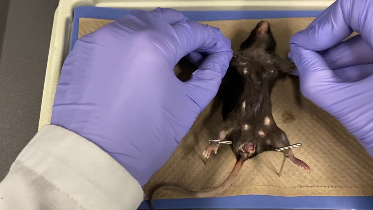
Image taken from the YouTube channel Unisciel , from the video titled Mouse dissection .
Unveiling the Anatomy and Physiology of Mus musculus for Research
Mus musculus, the common house mouse, stands as a cornerstone of modern biological and medical research. Its widespread use is not arbitrary; rather, it stems from a confluence of factors that render it an invaluable model organism for probing the intricacies of mammalian biology and disease.
Understanding the mouse's anatomy and physiology is paramount for accurate data interpretation, experimental design, and ultimately, for translating findings to human health. This section provides a foundational understanding of why the mouse holds such a prominent position in the scientific landscape.
A Legacy of Scientific Contribution
The use of mice in scientific inquiry dates back over a century, with early experiments focusing on genetics and basic biology. Over time, the mouse has become increasingly sophisticated as a model, mirroring advancements in molecular biology and genetic engineering.
The relatively short lifespan and ease of breeding, combined with its mammalian physiology, made the mouse an ideal candidate for studying complex biological processes across multiple generations in a condensed timeframe.
The Genetic Bridge: Mice and Humans
A key justification for the extensive use of mice lies in their remarkable genetic similarity to humans. Approximately 95% of human genes have a direct counterpart in the mouse genome. This high degree of conservation extends to many physiological pathways and disease mechanisms.
This genetic homology enables researchers to model human diseases in mice, investigate gene function, and test therapeutic interventions with a high degree of translational relevance. The ability to manipulate the mouse genome with precision further enhances its utility in studying the genetic basis of disease.
Broad Applications in Biomedical Science
The applications of mouse models span a vast spectrum of biological and medical research.
In disease research, mice are used to model a wide range of conditions, including cancer, cardiovascular disease, neurodegenerative disorders, and infectious diseases.
In drug development, mice serve as preclinical models to assess the efficacy and safety of novel therapeutic agents before human trials.
In basic biological studies, mice are employed to elucidate fundamental processes such as development, immunity, and aging.
The depth and breadth of mouse model applications underscore its indispensable role in advancing scientific knowledge and improving human health.
[Unveiling the Anatomy and Physiology of Mus musculus for Research Mus musculus, the common house mouse, stands as a cornerstone of modern biological and medical research. Its widespread use is not arbitrary; rather, it stems from a confluence of factors that render it an invaluable model organism for probing the intricacies of mammalian biology and...]
Foundational Disciplines: The Interwoven Fabric of Histology, Physiology, and Anatomy
A comprehensive understanding of Mus musculus, and indeed any biological system, rests upon the synergistic interplay of three foundational disciplines: histology, physiology, and anatomy. These are not discrete silos of knowledge; rather, they are interwoven strands that, when viewed collectively, provide a holistic perspective on the structure, function, and dynamic processes within the organism. Their combined insights are essential for interpreting experimental data and drawing meaningful conclusions in biomedical research.
Histology: Unveiling the Microscopic Architecture of Life
Histology, at its core, is the microscopic study of tissues. This discipline provides a critical bridge between the macroscopic world of anatomy and the molecular realm of biochemistry and genetics. It allows researchers to visualize the cellular organization of organs and tissues, revealing architectural details that are invisible to the naked eye.
The power of histology lies in its ability to reveal both normal tissue structure and pathological changes indicative of disease. Through careful examination of tissue samples, histologists can identify subtle alterations in cellular morphology, tissue organization, and the presence of abnormal cellular components.
The Art and Science of Staining
A cornerstone of histological analysis is the use of staining techniques. These techniques employ dyes that selectively bind to different cellular components, highlighting their structure and organization.
Perhaps the most widely used staining method is Hematoxylin and Eosin (H&E) staining. Hematoxylin, a basic dye, stains acidic structures like DNA and RNA a blue-purple color. Eosin, an acidic dye, stains basic structures like proteins and cytoplasm a pink-red color. This differential staining allows for clear visualization of cellular nuclei, cytoplasm, and extracellular matrix.
The Microscope: A Window into the Microscopic World
The microscope is the primary tool of the histologist. Various types of microscopes, including light microscopes and electron microscopes, provide different levels of magnification and resolution. Light microscopes are used for routine examination of stained tissue sections. Electron microscopes, with their much higher magnification capabilities, are used to visualize subcellular structures like organelles and protein complexes.
Physiology: Decoding the Functional Mechanisms
Physiology delves into the functional mechanisms that underpin life processes within Mus musculus.
It explores how organ systems function, how they interact, and how they are regulated to maintain internal stability. It is the study of how the "machine" works.
Homeostasis: The Cornerstone of Physiological Stability
A central concept in physiology is homeostasis. Homeostasis refers to the maintenance of a stable internal environment despite external fluctuations. This delicate balance is essential for optimal cellular function and overall organismal health. Physiological studies often focus on understanding the mechanisms that maintain homeostasis, such as feedback loops, hormonal regulation, and neural control.
Organ-System Interactions: A Symphony of Functions
Physiology also emphasizes the interconnectedness of organ systems. The cardiovascular, respiratory, digestive, and endocrine systems, among others, work in concert to ensure the survival and well-being of the organism.
Physiological studies often investigate how these systems interact and influence one another. For example, the cardiovascular and respiratory systems work together to deliver oxygen to tissues and remove carbon dioxide. Disruptions in one system can have cascading effects on others, highlighting the importance of understanding these complex interactions.
Anatomy: Mapping the Structural Landscape
Anatomy provides the structural framework for understanding the organism. It encompasses the study of both macroscopic and microscopic structures, from the overall organization of organ systems to the intricate details of individual cells and tissues.
Systemic Exploration: The Anatomist's Approach
Anatomists systematically explore the body, identifying and describing the location, size, shape, and relationships of various organs and tissues. This detailed mapping is essential for understanding how the body is organized and how its different parts work together.
Comparative Anatomy: Insights from Evolutionary History
Comparative anatomy examines the similarities and differences in anatomical structures across different species. By comparing the anatomy of Mus musculus to that of other mammals, including humans, researchers can gain insights into evolutionary relationships and the functional significance of different anatomical features. This comparative approach is particularly valuable for understanding the evolution of disease mechanisms and for developing more effective treatments.
[[Unveiling the Anatomy and Physiology of Mus musculus for Research Mus musculus, the common house mouse, stands as a cornerstone of modern biological and medical research. Its widespread use is not arbitrary; rather, it stems from a confluence of factors that render it an invaluable model organism for probing the intricacies of mammalian biology an...]]
A Deep Dive into Anatomical Systems: Structure and Function
To harness the full potential of Mus musculus as a research model, a meticulous understanding of its anatomy is paramount. This section embarks on a detailed exploration of the major anatomical systems, elucidating their structural components and functional roles within the organism. We delve into each system, providing a comprehensive overview necessary for researchers seeking to contextualize experimental results and design targeted investigations.
The Skeletal System: Foundation of Form and Movement
The skeletal system provides the structural framework of the mouse, offering support, protection, and enabling locomotion.
The cranium, or skull, serves as a robust protective vault for the delicate brain, safeguarding it from physical trauma.
The vertebral column, composed of individual vertebrae, provides flexible support for the body and houses the spinal cord, a critical component of the nervous system.
The rib cage encases and protects the thoracic organs, including the heart and lungs, from external forces.
The forelimbs and hindlimbs, with their intricate arrangement of bones, facilitate movement and interaction with the environment. Key bones like the femur, tibia, and humerus are essential for weight-bearing and propulsion.
The Muscular System: Orchestrating Movement
The muscular system is responsible for generating movement, maintaining posture, and producing heat. Skeletal muscles, attached to the skeleton via tendons, contract to produce voluntary movements.
These muscles are composed of muscle fibers, specialized cells containing contractile proteins. The coordinated action of multiple muscles allows for a wide range of movements, from fine motor skills to powerful locomotion.
The Nervous System: Command and Control Center
The nervous system acts as the body's command and control center, receiving sensory information, processing it, and coordinating responses.
The brain, comprising the cerebrum, cerebellum, and brainstem, is the central processing unit, responsible for higher-level functions such as learning, memory, and decision-making.
The spinal cord serves as a neural pathway, transmitting signals between the brain and the peripheral nervous system.
Peripheral nerves extend from the central nervous system to innervate muscles, glands, and sensory organs throughout the body. Neurons, the fundamental cellular units of the nervous system, transmit electrical and chemical signals.
The Cardiovascular System: Delivering Life's Essentials
The cardiovascular system is responsible for circulating blood throughout the body, delivering oxygen and nutrients to tissues and removing waste products.
The heart acts as a pump, propelling blood through a network of blood vessels, including arteries, veins, and capillaries.
Arteries carry oxygenated blood away from the heart, while veins return deoxygenated blood to the heart. Capillaries, the smallest blood vessels, facilitate the exchange of gases, nutrients, and waste products between the blood and surrounding tissues. Blood, composed of red blood cells, white blood cells, and plasma, is the circulatory fluid that carries these essential substances.
The Respiratory System: Facilitating Gas Exchange
The respiratory system is responsible for gas exchange, taking in oxygen from the air and expelling carbon dioxide.
The lungs are the primary organs of respiration, where oxygen and carbon dioxide are exchanged between the air and the blood.
The trachea and bronchi form the airway, conducting air to and from the lungs. Alveoli, tiny air sacs within the lungs, provide a large surface area for gas exchange.
The diaphragm, a dome-shaped muscle located at the base of the thoracic cavity, plays a crucial role in breathing by contracting and relaxing to change the volume of the chest cavity.
The Digestive System: Processing and Absorbing Nutrients
The digestive system is responsible for breaking down food, absorbing nutrients, and eliminating waste products.
Food enters the body through the mouth, where it is mechanically broken down and mixed with saliva.
The esophagus transports food from the mouth to the stomach, where initial digestion occurs.
The small intestine is the primary site of nutrient absorption, while the large intestine absorbs water and forms waste products.
The liver plays a crucial role in metabolism, detoxifying harmful substances and producing bile. The pancreas secretes enzymes that aid in digestion and hormones that regulate blood sugar levels.
The Endocrine System: Hormonal Regulation
The endocrine system regulates various bodily functions through the secretion of hormones.
The pituitary gland, often referred to as the "master gland," controls the activity of other endocrine glands.
The thyroid gland regulates metabolism, while the adrenal glands mediate the stress response and influence metabolism.
The pancreas (specifically the islets of Langerhans) produces insulin and glucagon, which regulate blood sugar levels. The gonads (testes in males and ovaries in females) produce sex hormones that regulate reproductive function.
The Reproductive System: Perpetuating the Species
The reproductive system is responsible for procreation.
In males, the testes produce sperm, which mature in the epididymis and are transported through the vas deferens. The prostate gland and seminal vesicles contribute fluids to semen.
In females, the ovaries produce eggs, which travel through the fallopian tubes to the uterus, where fetal development occurs. The vagina is the canal connecting the uterus to the external environment.
The Integumentary System: Protection and Sensory Input
The integumentary system, consisting of the skin, hair, and associated structures, provides a protective barrier against the external environment.
The skin, composed of the epidermis and dermis, protects against infection, dehydration, and physical damage. Hair provides insulation and sensory input. Claws, keratinous structures on the digits, aid in locomotion and defense.
Sebaceous glands secrete sebum, an oily substance that lubricates the skin and hair. Sweat glands help regulate body temperature through perspiration.
The Sensory Organs: Perceiving the World
The sensory organs allow the mouse to perceive its environment.
The eyes provide visual perception, allowing the mouse to navigate its surroundings and detect predators.
The ears are responsible for auditory perception and balance. The nose detects odors, playing a crucial role in finding food and identifying other mice.
The tongue detects tastes, while the skin contains touch receptors that provide tactile information. These sensory inputs are essential for the mouse's survival and interaction with its environment.
Essential Experimental Tools and Techniques for Mus musculus Studies
Following our comprehensive anatomical overview, it is crucial to examine the tools and techniques that empower researchers to dissect, visualize, and analyze the intricate biology of Mus musculus. Mastering these methodologies is paramount for generating robust and reproducible data.
Dissection Tools: Precision Instruments for Anatomical Exploration
Dissection forms the bedrock of anatomical investigation, requiring a specialized toolkit to navigate the delicate structures of the mouse. High-quality stainless steel instruments are essential to minimize tissue damage and ensure accurate observation.
A basic dissection kit typically includes:
- Scalpels: For making precise incisions. Blades of varying sizes provide versatility.
- Forceps: For grasping and manipulating tissues. Fine-tipped forceps are crucial for delicate structures.
- Scissors: For cutting tissues and vessels. Micro-scissors are essential for intricate dissections.
- Probes: For blunt dissection and tissue manipulation.
- Dissecting Needles: For teasing apart tissues and identifying structures.
Beyond the basic tools, specialized instruments such as micro-dissectors and surgical microscopes may be required for advanced anatomical studies. Proper sterilization of dissection tools is paramount to prevent contamination and maintain the integrity of the sample.
Imaging Techniques: Visualizing the Internal Landscape
Imaging technologies provide non-invasive or minimally invasive means of visualizing the internal anatomy and physiology of Mus musculus. These modalities offer complementary perspectives, enabling researchers to examine structures in vivo or ex vivo.
X-ray Radiography: A Foundation for Skeletal Imaging
X-ray radiography, utilizing electromagnetic radiation, remains a fundamental technique for visualizing bone structures and detecting abnormalities such as fractures or tumors. Its relative simplicity and affordability make it a widely accessible imaging modality. However, its limited soft tissue contrast restricts its utility for detailed anatomical studies of organs and other soft tissues.
Magnetic Resonance Imaging (MRI): High-Resolution Soft Tissue Visualization
MRI employs powerful magnets and radio waves to generate high-resolution images of soft tissues. It excels in differentiating various tissue types and visualizing subtle anatomical details within organs, the brain, and other structures. MRI is particularly valuable for longitudinal studies, allowing researchers to track changes in tissue structure over time.
Computed Tomography (CT) Scans: Detailed Cross-Sectional Imaging
CT scans, also known as CAT scans, utilize X-rays to create detailed cross-sectional images of the body. These images can be reconstructed into three-dimensional models, providing comprehensive anatomical information. CT imaging is particularly useful for visualizing bone structures, detecting tumors, and assessing vascular anatomy.
Ultrasound Imaging: Real-Time, Non-Invasive Visualization
Ultrasound imaging utilizes high-frequency sound waves to visualize internal structures in real-time. Its non-invasive nature and portability make it a valuable tool for studying cardiovascular function, monitoring fetal development, and guiding interventional procedures. However, image quality can be affected by factors such as tissue density and operator skill.
Immunohistochemistry: Mapping Protein Expression
Immunohistochemistry (IHC) is a powerful technique for visualizing the expression and localization of specific proteins within tissue sections. It involves using antibodies that selectively bind to target proteins, followed by a detection system that allows for visualization under a microscope.
IHC is essential for:
- Identifying specific cell types within tissues.
- Studying protein expression patterns in different tissues or disease states.
- Determining the subcellular localization of proteins.
- Validating the results of gene expression studies.
Careful optimization of IHC protocols is crucial to ensure accurate and reliable results. Factors such as antibody selection, antigen retrieval methods, and blocking steps can significantly impact the quality of staining.
The Mouse as a Model Organism: Genetic Context and Strain Diversity
Following our comprehensive exploration of the anatomy and physiology of Mus musculus, it's critical to understand the genetic underpinnings that make it such a valuable research tool. This section delves into the genetic characteristics of the mouse, the diverse strains available, and the powerful applications of genetically engineered mouse models in biomedical research.
Mus musculus: A Cornerstone of Modern Research
Mus musculus, the common house mouse, occupies a unique and indispensable position in the landscape of biological and medical research. Its relatively short lifespan, ease of breeding, and physiological similarities to humans make it an ideal model for studying a wide range of diseases and biological processes.
Furthermore, the mouse genome has been fully sequenced and is remarkably similar to the human genome, enabling researchers to translate findings from mouse models to human health more effectively. The extensive genetic tools and resources available for mice are unparalleled, making them a preferred choice for investigating complex biological questions.
Strain Diversity: Tailoring the Model to the Question
The genetic diversity within Mus musculus is a significant asset for researchers. Different inbred strains exhibit unique characteristics, making them suitable for modeling specific diseases or studying particular biological pathways. Understanding the properties of these strains is crucial for selecting the most appropriate model for a given research question.
C57BL/6 (Black 6): The Workhorse of Research
The C57BL/6 strain is arguably the most widely used inbred mouse strain in research. Known for its robustness, relatively low incidence of spontaneous tumors, and well-characterized genome, C57BL/6 serves as a background strain for many genetically modified models. It is frequently used in studies of immunology, neuroscience, and cardiovascular disease.
BALB/c: An Immunological Standout
BALB/c mice are characterized by their albino coat color and heightened susceptibility to certain types of cancer. They are particularly valuable in immunology research, as they exhibit a strong Th2 immune response, making them useful models for studying allergy and asthma.
FVB/N: Transgenic Powerhouse
FVB/N mice are known for their large pronuclei, which makes them highly amenable to pronuclear microinjection, a technique used to create transgenic mice. This strain is often used as a background for generating transgenic models and is valuable for studying developmental biology and cancer.
Swiss Webster: A General Purpose Model
Swiss Webster mice are an outbred strain, meaning they possess a higher degree of genetic diversity than inbred strains. This genetic variability can be both an advantage and a disadvantage. Swiss Webster mice are often used as a general-purpose model for toxicology studies and drug development.
NOD (Non-Obese Diabetic): Modeling Type 1 Diabetes
NOD mice spontaneously develop autoimmune type 1 diabetes, making them an invaluable model for studying the pathogenesis of this disease. These mice are used to investigate the genetic and environmental factors that contribute to the development of type 1 diabetes and to test potential therapies.
SCID (Severe Combined Immunodeficiency): A Window into the Immune System
SCID mice lack functional T and B cells, rendering them severely immunodeficient. These mice are used as recipients for xenografts, allowing researchers to study human tumors and immune responses in vivo.
Genetically Engineered Mouse Models: Precision Tools for Disease Research
The ability to manipulate the mouse genome has revolutionized biomedical research. Genetically engineered mouse models (GEMMs) allow researchers to study the effects of specific genes on development, physiology, and disease progression. These models are powerful tools for understanding the molecular mechanisms underlying human diseases and for developing novel therapies.
Transgenic Mice: Adding New Genes
Transgenic mice carry a foreign gene (transgene) that has been artificially inserted into their genome. This technique allows researchers to study the effects of overexpressing a particular gene or to express a gene in a specific tissue or cell type. Transgenic mice have been used to model a wide range of diseases, including cancer, neurodegenerative disorders, and cardiovascular disease.
Knockout Mice: Silencing Genes to Uncover Function
Knockout mice have a specific gene that has been inactivated or "knocked out." By studying the effects of gene deletion, researchers can determine the function of the missing gene and its role in various biological processes. Knockout mice are invaluable for understanding the genetic basis of disease and for identifying potential drug targets.
Conditional Knockout Mice: Spatiotemporal Precision
Conditional knockout mice allow researchers to inactivate a gene in a specific tissue or at a specific time point. This level of control is achieved by using the Cre-loxP system, which allows for targeted gene deletion. Conditional knockout mice are particularly useful for studying genes that have essential functions during development or in multiple tissues.
Expertise and Collaboration: The Role of Anatomists and Veterinarians
The Mouse as a Model Organism: Genetic Context and Strain Diversity Following our comprehensive exploration of the anatomy and physiology of Mus musculus, it's critical to understand the genetic underpinnings that make it such a valuable research tool. This section delves into the genetic characteristics of the mouse, the diverse strains available, and the pivotal roles of anatomists and laboratory animal veterinarians in maximizing the scientific validity and ethical treatment of these animals. Their expertise is indispensable to ensure research integrity and animal well-being.
The Indispensable Anatomist: A Foundation of Knowledge
Anatomists bring specialized expertise to the study of Mus musculus, providing a critical foundation for research. They possess an in-depth understanding of the structural organization of the mouse body.
This includes both macroscopic and microscopic levels. Their knowledge extends to the intricate relationships between different organ systems.
Anatomists contribute significantly to experimental design, ensuring proper techniques are used for sample collection and analysis. They are crucial for interpreting morphological data.
Without a deep understanding of normal anatomy, it is impossible to accurately identify and interpret pathological changes.
Moreover, anatomists are instrumental in training researchers. They provide essential guidance on dissection techniques and anatomical landmarks. Their insights are critical for accurate phenotyping.
Veterinarians Specializing in Laboratory Animal Medicine: Guardians of Welfare and Scientific Validity
Veterinarians specializing in laboratory animal medicine are indispensable for ensuring the health and welfare of research mice. They play a multifaceted role.
Their primary responsibility is to prevent and treat diseases that could compromise the scientific validity of studies. They also monitor the overall health and well-being of the animals.
This involves implementing rigorous health surveillance programs. It also involves providing expert veterinary care when needed.
Veterinarians ensure that animal handling procedures are humane and ethical, adhering to the highest standards of animal welfare. They are vital in protocol review.
Their contributions ensure compliance with ethical guidelines and regulations.
Refinement, Reduction, and Replacement: The Core Principles
Laboratory animal veterinarians are champions of the "3Rs" – Refinement, Reduction, and Replacement.
Refinement involves improving husbandry and experimental procedures to minimize pain and distress. Reduction aims to minimize the number of animals used in research while still achieving statistically significant results.
Replacement seeks to replace animal models with in vitro or in silico methods whenever possible. Veterinarians actively promote these principles.
They work to integrate them into research practices.
Collaborative Synergy: Maximizing Impact
The most impactful research involving Mus musculus occurs when anatomists and laboratory animal veterinarians collaborate closely.
This interdisciplinary approach ensures that studies are both scientifically rigorous and ethically sound.
Anatomists provide the detailed anatomical knowledge necessary for accurate interpretation of experimental results. Veterinarians ensure the animals are healthy and free from confounding factors.
Together, they contribute to the generation of reliable and reproducible data, advancing scientific knowledge while upholding the highest standards of animal welfare. Their collaborative expertise is essential.
It is truly essential for the responsible and productive use of Mus musculus in research.
Video: Mice Anatomy: The Ultimate Guide to Mouse Biology
FAQs: Mice Anatomy - The Ultimate Guide
What specific areas of mice anatomy does the guide cover?
The guide covers all major organ systems, including skeletal, muscular, nervous, circulatory, respiratory, digestive, and reproductive systems. It also explores external features and unique aspects of mice anatomy.
Why is understanding mice anatomy important?
Understanding mice anatomy is crucial for researchers and veterinarians. It helps interpret research data, diagnose illnesses, and perform procedures accurately. Good understanding of mice anatomy aids in the ethical treatment and care of laboratory mice.
Does the guide detail differences between mouse strains?
Yes, the guide highlights key anatomical variations between common laboratory mouse strains. Understanding these subtle differences in mice anatomy is important for experiment design.
What level of detail does the guide provide on microscopic anatomy?
The guide includes detailed information on microscopic anatomy, or histology, of various mouse tissues and organs. It covers cellular structures and their functions, crucial for research involving mice anatomy at the cellular level.
So, there you have it! Hopefully, this dive into mice anatomy has shed some light on these fascinating little creatures. Whether you're a student, researcher, or just plain curious, understanding the intricacies of mouse biology is truly remarkable. Now, go forth and appreciate the amazing complexity packed into these tiny mammals!
