Clinical Attachment Loss: Stages You Need to Know Now!
Gingival recession, a common precursor to clinical attachment loss, often signals the initial stages of periodontal disease. This deterioration process can be thoroughly assessed utilizing a periodontal probe, an essential instrument in dental diagnostics. The American Academy of Periodontology (AAP) emphasizes the importance of early detection and intervention to prevent progression. Understanding the various stages of clinical attachment loss is paramount for dentists, hygienists, and even patients seeking to proactively manage their oral health and potentially avoid more invasive treatments recommended by experts like Dr. Paul Keyes.

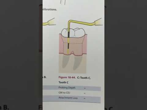
Image taken from the YouTube channel Dentiscope , from the video titled Dentiscope | HOW TO DETERMINE CLINICAL ATTACHMENT LOSS .
The health of our gums and the supporting structures of our teeth—collectively known as the periodontium—is often an overlooked aspect of overall well-being.
Yet, periodontal health is critical, and when compromised, it can lead to a condition known as Clinical Attachment Loss (CAL).
This silent but progressive deterioration can have significant consequences for your oral health and, potentially, your overall health.
The insidious nature of CAL is that it often goes unnoticed in its early stages, allowing the damage to progress gradually.
Early detection and intervention are paramount to halting its advancement and preserving the integrity of your smile.
This article aims to shed light on Clinical Attachment Loss, exploring its definition, significance, and the distinct stages through which it progresses.
Our primary goal is to empower you with the knowledge to understand your oral health better, recognize potential problems early, and take proactive steps to maintain a healthy smile for years to come.
What Exactly is Clinical Attachment Loss (CAL)?
Clinical Attachment Loss (CAL) refers to the irreversible destruction of the tissues that hold your teeth in place.
These tissues include the gingiva (gums), the periodontal ligament (which connects the tooth to the bone), the cementum (the outer layer of the tooth root), and the alveolar bone (the bone that surrounds and supports the teeth).
CAL is typically measured as the distance from the cementoenamel junction (CEJ), where the enamel of the crown meets the cementum of the root, to the most apical point of attachment of the periodontal tissues.
A greater distance indicates more severe attachment loss.
Unlike gingivitis, which is characterized by inflammation of the gums and is often reversible with proper oral hygiene, CAL signifies that the damage has extended beyond the gums and into the supporting structures of the teeth.
This damage is permanent, making early detection and prevention even more crucial.
The Critical Importance of Early Detection and Intervention
The progression of Clinical Attachment Loss can have far-reaching implications for your oral health.
As the supporting tissues deteriorate, teeth can become loose, shift in position, or even be lost entirely.
Beyond tooth loss, periodontal disease, of which CAL is a key indicator, has been linked to several systemic health conditions, including cardiovascular disease, diabetes, and respiratory infections.
This connection underscores the importance of maintaining good periodontal health for your overall well-being.
Early detection of CAL allows for timely intervention, which can help to slow or even halt the progression of the disease.
Treatment options range from non-surgical procedures, such as scaling and root planing (deep cleaning), to surgical interventions aimed at regenerating lost tissue.
The earlier CAL is detected and treated, the more successful the treatment is likely to be, and the better the long-term prognosis for your teeth and gums.
Empowering You to Understand Your Oral Health
This article is designed to be a comprehensive guide to understanding Clinical Attachment Loss.
We will break down the complex topic into easily digestible information, focusing on the different stages of CAL and the key indicators associated with each stage.
By understanding these stages, you will be better equipped to recognize potential warning signs, ask informed questions during dental appointments, and take proactive steps to protect your periodontal health.
Ultimately, our goal is to empower you to take control of your oral health and work in partnership with your dentist or periodontist to maintain a healthy, confident smile for life.
The Foundation: Understanding Periodontal Health
Before we delve deeper into Clinical Attachment Loss, it's essential to establish a firm understanding of what constitutes a healthy periodontium.
The periodontium is essentially the foundation that supports your teeth, ensuring their stability and proper function.
Think of it as the scaffolding that holds a building upright; when this foundation is compromised, the entire structure is at risk.
Let's explore the key components of this vital system: the gingiva, periodontal ligament, cementum, and alveolar bone.
Components of a Healthy Periodontium
The periodontium isn't a single entity, but rather a team of specialized tissues working in harmony.
Understanding each component's role is critical to appreciating the overall health of your gums and teeth.
-
Gingiva (Gums): The gingiva, or gums, is the visible soft tissue that surrounds the necks of the teeth and covers the alveolar bone.
Healthy gingiva is typically pink, firm, and exhibits a stippled texture (similar to an orange peel). Its primary function is to protect the underlying tissues from bacterial invasion and physical trauma.
-
Periodontal Ligament (PDL): The periodontal ligament is a complex network of connective tissue fibers that attach the tooth root to the alveolar bone.
It acts as a shock absorber, cushioning the tooth from the forces of chewing and biting.
The PDL also contains cells that are essential for the maintenance and repair of the surrounding tissues.
-
Cementum: Cementum is a thin, calcified layer that covers the root of the tooth.
Its primary function is to provide an attachment surface for the periodontal ligament fibers.
Cementum is softer than enamel and dentin, making it susceptible to abrasion if exposed due to gingival recession.
-
Alveolar Bone: The alveolar bone is the bone that surrounds and supports the teeth within the jawbone.
It contains sockets, called alveoli, in which the teeth are anchored.
The alveolar bone is constantly being remodeled in response to forces placed upon the teeth. It is also susceptible to resorption in the presence of periodontal disease.
The Indispensable Role of Oral Hygiene
Maintaining excellent oral hygiene is paramount to preserving the health of your periodontium.
Regular brushing, flossing, and professional dental cleanings help remove plaque and calculus (tartar), the primary culprits behind periodontal disease.
Plaque is a sticky film of bacteria that constantly forms on your teeth. If not removed, it can harden into calculus, which is much more difficult to eliminate.
The bacteria in plaque and calculus produce toxins that irritate the gums, leading to inflammation (gingivitis).
If gingivitis is left untreated, it can progress to periodontitis, characterized by Clinical Attachment Loss and eventual tooth loss.
Therefore, a consistent and thorough oral hygiene routine is your first line of defense against periodontal disease, safeguarding the health and integrity of your entire periodontium.
What is Clinical Attachment Loss (CAL)?
Having explored the foundation of a healthy periodontium, the next logical step is to understand what happens when that foundation begins to crumble.
Clinical Attachment Loss (CAL) represents a significant and irreversible deterioration of the tissues that support your teeth.
It's a critical indicator of periodontal disease progression and a key factor in determining the long-term prognosis of your teeth.
Defining Clinical Attachment Loss
Clinical Attachment Loss is defined as the distance from the cementoenamel junction (CEJ) to the base of the periodontal pocket.
In simpler terms, it's a measurement of how much the gums and bone have receded away from the crown of the tooth due to periodontal disease.
This recession leads to a loss of the connective tissue attachment that anchors the tooth in place.
The more attachment that is lost, the less support the tooth has, and the greater the risk of tooth mobility and eventual tooth loss.
CAL vs. Gingivitis: Understanding the Difference
It's essential to differentiate Clinical Attachment Loss from gingivitis.
Gingivitis is an inflammation of the gums that is usually reversible with good oral hygiene.
The key distinction is that gingivitis does not involve any loss of attachment or bone.
In contrast, Clinical Attachment Loss always indicates irreversible damage to the periodontal tissues.
While gingivitis can be a precursor to periodontitis, it doesn't always progress to that stage.
However, once Clinical Attachment Loss occurs, the disease has progressed to periodontitis.
This makes early detection and treatment crucial to prevent further damage.
The Role of Plaque and Calculus
The primary culprit behind periodontitis and Clinical Attachment Loss is dental plaque, a sticky film of bacteria that constantly forms on our teeth.
When plaque is not removed effectively through brushing and flossing, it can harden into calculus, also known as tartar.
Calculus provides a rough surface that further promotes plaque accumulation and makes it more difficult to remove.
The bacteria in plaque produce toxins that irritate the gums, triggering an inflammatory response.
This chronic inflammation leads to the destruction of the periodontal tissues, resulting in Clinical Attachment Loss.
The longer plaque and calculus remain on the teeth, the greater the risk of developing periodontitis and experiencing irreversible Clinical Attachment Loss.
With a clearer understanding of how CAL is defined and differentiated from other gum conditions, it's time to delve into the progression of this disease. The insidious nature of periodontal disease lies in its gradual development, making early detection paramount.
The Four Stages of Clinical Attachment Loss: A Detailed Breakdown
Periodontal disease, and consequently Clinical Attachment Loss, doesn't occur overnight. It progresses through distinct stages, each characterized by specific clinical signs and symptoms. Understanding these stages is vital for both dental professionals and individuals seeking to proactively manage their oral health. Accurate staging allows for tailored treatment plans and helps predict the long-term prognosis of affected teeth.
Stage 1: Initial Stage (Early Periodontitis)
This initial stage often goes unnoticed, as the symptoms can be subtle. However, it's a critical window for intervention.
Early signs include:
- Slight Gingival Recession: The gum line may start to pull away from the teeth, exposing a small portion of the root surface.
- Minimal Alveolar Bone Loss: Radiographs (X-rays) may reveal slight bone loss around the teeth. This bone loss is often localized.
Pocket Depth and Bleeding on Probing
Pocket Depth measurements typically range from 1-2mm at this stage. While this might seem insignificant, it indicates the beginning of pocket formation.
Bleeding on Probing (BOP) is a key indicator. Even with shallow pockets, the gums are inflamed, and bleeding easily occurs when gently probed. BOP signals active inflammation and highlights the need for improved oral hygiene. The absence of bleeding is a good sign, but it is not a guarantee that the site is healthy.
Stage 2: Moderate Periodontitis
As the disease progresses, the signs and symptoms become more noticeable. Stage 2 represents a more significant threat to the supporting structures of the teeth.
Key characteristics include:
- Increased Clinical Attachment Loss: There is a measurable loss of attachment, indicating the disease is actively destroying the tissues that hold the teeth in place.
- Moderate Alveolar Bone Loss: Radiographs clearly show more bone loss compared to Stage 1, affecting a larger area around the tooth roots.
Pocket Depth and Tooth Mobility
Deeper Pocket Depth measurements are typically observed, ranging from 3-4mm. These deeper pockets provide an environment for bacteria to thrive, further exacerbating the inflammatory process.
Possible Increase in Tooth Mobility: The affected teeth may start to exhibit slight mobility. This is because the supporting bone has been reduced, weakening their anchorage.
Stage 3: Severe Periodontitis
Stage 3 signifies a substantial compromise in the periodontal support of the teeth. The damage is significant, and without intervention, tooth loss is a real possibility.
Hallmarks of this stage include:
- Significant Clinical Attachment Loss: The loss of attachment is extensive, severely impacting the stability of the teeth.
- Advanced Alveolar Bone Loss: Radiographs reveal significant bone loss, often extending to the middle third of the tooth root or beyond.
Pocket Depth, Tooth Mobility, and Furcation Involvement
Deep Pocket Depth: Pocket depths are typically 6mm or greater. These deep pockets are extremely difficult to clean, even with professional intervention.
Increased Tooth Mobility: Teeth exhibit noticeable mobility, making chewing uncomfortable. The risk of tooth loss is significantly elevated.
May involve Furcation Involvement: In multi-rooted teeth (molars), the bone between the roots, called the furcation, may be affected. This furcation involvement further compromises the tooth's stability.
Stage 4: Advanced Periodontitis
This is the most advanced and destructive stage of periodontal disease. It represents a severe threat to the entire dentition.
Characteristics include:
- Severe Clinical Attachment Loss: The loss of attachment is extreme, leaving very little support for the affected teeth.
- Extensive Alveolar Bone Loss: Radiographs show severe bone loss, often extending to the apex (tip) of the tooth root.
Tooth Mobility and Treatment Complexity
Significant Tooth Mobility and Potential Tooth Loss: Teeth exhibit extreme mobility and may be on the verge of falling out. Tooth loss is highly likely without aggressive treatment.
Complex Periodontal Issues Requiring Comprehensive Treatment: Stage 4 often involves multiple teeth and complex treatment needs. Extraction of hopeless teeth, bone grafting, and extensive restorative work may be necessary to restore function and aesthetics.
With a clearer understanding of how CAL is defined and differentiated from other gum conditions, it's time to delve into the progression of this disease. The insidious nature of periodontal disease lies in its gradual development, making early detection paramount.
Diagnosis and Assessment: Identifying CAL
The accurate diagnosis of Clinical Attachment Loss (CAL) is a critical step in managing periodontal disease and preventing further damage to the supporting structures of the teeth.
It requires a comprehensive evaluation by a qualified dental professional, either a general dentist or, in more complex cases, a periodontist. This process involves a combination of clinical examination and radiographic assessment to determine the extent and severity of the condition.
The Roles of Dentists and Periodontists
Both general dentists and periodontists play essential roles in identifying CAL. General dentists are often the first line of defense, screening for early signs of periodontal disease during routine check-ups.
They can detect gingival inflammation, bleeding, and early pocket formation, prompting further investigation if necessary.
Periodontists, on the other hand, are specialists in the diagnosis, treatment, and prevention of periodontal diseases.
They possess advanced training and expertise in assessing complex cases of CAL and developing tailored treatment plans.
A referral to a periodontist may be warranted when the condition is severe, rapidly progressing, or unresponsive to initial treatment.
Periodontal Probing: Measuring Pocket Depth and Attachment Loss
Periodontal probing is a fundamental diagnostic procedure used to measure the depth of the gingival sulcus or periodontal pocket.
This measurement is taken using a calibrated periodontal probe, a slender instrument marked with millimeter increments.
The probe is gently inserted between the tooth and the gum tissue to assess the distance from the gingival margin to the base of the pocket.
In healthy gums, the pocket depth typically ranges from 1 to 3 millimeters. Increased pocket depth indicates the presence of periodontal disease and the formation of a deeper space where bacteria can accumulate.
In addition to pocket depth, periodontal probing also allows the clinician to assess Clinical Attachment Loss (CAL) directly.
CAL represents the distance from the cementoenamel junction (CEJ) – the point where the enamel and cementum meet on the tooth – to the base of the pocket.
This measurement provides a more accurate indication of the amount of tissue destruction that has occurred, as it accounts for gingival recession.
Radiographic Evaluation: Assessing Alveolar Bone Loss
Radiographs, or X-rays, are an indispensable tool in the diagnosis and assessment of CAL. They provide a visual representation of the alveolar bone, the bone that supports the teeth.
Radiographs can reveal the extent and pattern of bone loss associated with periodontal disease, which is often not visible during a clinical examination alone.
Different types of radiographs, such as periapical and bitewing X-rays, may be used to assess the bone levels around the teeth.
The dentist or periodontist will carefully examine the radiographs to identify any areas of bone loss, paying attention to the shape, depth, and distribution of the bone defects.
The amount of bone loss is typically categorized as mild, moderate, or severe, depending on the percentage of root length affected.
Radiographic evaluation is also crucial for detecting other factors that may contribute to CAL, such as:
- Furcation involvement: Bone loss in the area where the roots of a multi-rooted tooth diverge.
- Periapical lesions: Infections or inflammations at the tip of the tooth root.
- Root fractures: Cracks or breaks in the tooth root.
By combining clinical examination with radiographic assessment, dental professionals can accurately diagnose CAL, determine the severity of periodontal disease, and develop appropriate treatment plans to restore and maintain periodontal health.
With a clearer understanding of how CAL is defined and differentiated from other gum conditions, it's time to delve into the progression of this disease. The insidious nature of periodontal disease lies in its gradual development, making early detection paramount.
Treatment Options: Restoring Periodontal Health
The management of Clinical Attachment Loss (CAL) requires a multi-faceted approach, focusing on halting disease progression and, where possible, restoring lost tissue. The specific treatment plan is tailored to the stage and severity of the CAL, along with individual patient factors. Effective treatment not only addresses the immediate problem but also equips the patient with the knowledge and tools for long-term periodontal health.
Non-Surgical Interventions: The First Line of Defense
In the early to moderate stages of CAL, non-surgical treatments are often the initial approach.
Scaling and Root Planing (SRP): A Deep Cleaning
Scaling and Root Planing (SRP), often referred to as deep cleaning, is the cornerstone of non-surgical periodontal therapy. This procedure involves meticulously removing plaque, calculus (tartar), and bacterial toxins from the tooth surfaces, both above and below the gum line.
Scaling removes the deposits, while root planing smooths the root surfaces to discourage further bacterial adhesion and promote healing. Local anesthesia is typically used to ensure patient comfort during the procedure.
Adjunctive Therapies
Antimicrobial mouth rinses, such as chlorhexidine, may be prescribed to further reduce the bacterial load in the mouth. Systemic antibiotics are sometimes used in conjunction with SRP in cases of aggressive or refractory periodontitis.
Surgical Interventions: Addressing Advanced Cases
When non-surgical treatments are insufficient to control the disease, or in cases of advanced CAL, periodontal surgery may be necessary. These procedures aim to reduce pocket depth, regenerate lost tissue, and improve access for cleaning.
Pocket Reduction Surgery (Flap Surgery)
This procedure involves lifting the gums away from the teeth to allow for thorough cleaning of the root surfaces and underlying bone. In some cases, the bone may be reshaped to eliminate pockets and create a more favorable environment for healing. The gums are then sutured back into place, often at a position that reduces pocket depth.
Guided Tissue Regeneration (GTR)
Guided Tissue Regeneration (GTR) is a surgical technique used to regenerate lost periodontal tissues, including bone and ligaments. A membrane is placed between the gum and bone to prevent the gum tissue from growing into the space where bone and ligament should be regenerating. This allows the slower-growing bone and ligament cells to repopulate the area, leading to tissue regeneration.
Bone Grafting
When significant bone loss has occurred, bone grafting may be used to restore bone support around the teeth. Bone grafts can be obtained from various sources, including the patient's own body (autograft), a donor (allograft), or synthetic materials. The graft material provides a scaffold for new bone to grow, improving tooth stability and long-term prognosis.
Periodontal Plastic Surgery
These procedures address gum recession and improve the aesthetics of the smile. Techniques such as gum grafting can be used to cover exposed tooth roots, protect them from sensitivity and decay, and create a more natural-looking gum line.
Managing Contributing Factors: A Holistic Approach
Successful periodontal treatment requires addressing not only the disease itself but also the contributing factors that may exacerbate it.
Smoking Cessation
Smoking is a major risk factor for periodontal disease, impairing healing and increasing the likelihood of disease progression. Smoking cessation is crucial for successful treatment outcomes and long-term periodontal health.
Diabetes Management
Uncontrolled diabetes can increase the risk and severity of periodontal disease. Effective diabetes management is essential for controlling periodontal inflammation and promoting healing.
Stress Reduction
Stress can weaken the immune system and contribute to inflammation. Implementing stress reduction techniques can support overall health and improve the body's ability to fight infection.
The Role of the Periodontist
Periodontists are dental specialists with advanced training in the diagnosis, treatment, and prevention of periodontal diseases. They possess the expertise and skills to manage complex cases of CAL and perform advanced surgical procedures. A referral to a periodontist may be warranted when the condition is severe, rapidly progressing, or unresponsive to initial treatment.
In conclusion, restoring periodontal health after Clinical Attachment Loss requires a tailored approach, combining professional treatments with diligent self-care and management of contributing factors. With appropriate intervention and ongoing maintenance, it is possible to halt disease progression and preserve the dentition.
With a clearer understanding of how CAL is defined and differentiated from other gum conditions, it's time to delve into the progression of this disease. The insidious nature of periodontal disease lies in its gradual development, making early detection paramount.
Prevention and Maintenance: A Lifelong Commitment
The battle against Clinical Attachment Loss (CAL) isn't a sprint; it's a marathon. Even after successful treatment, the threat of recurrence looms large. Therefore, prevention and meticulous maintenance become absolutely critical, transforming from mere recommendations into a lifelong commitment to oral health. These are the cornerstones of preventing disease progression and ensuring long-term stability.
The Power of Daily Oral Hygiene
Excellent oral hygiene practices form the bedrock of any successful periodontal maintenance strategy. These practices directly combat the primary culprit behind CAL: bacterial plaque. Consistency and technique are equally vital, ensuring thorough removal of plaque from all tooth surfaces and along the gumline.
Brushing: The Foundation
Brushing at least twice a day with fluoride toothpaste is the starting point. The key lies in using proper technique. Gentle, circular motions or the Bass technique (angling the bristles towards the gumline) are recommended. Avoid aggressive scrubbing, which can damage gums and tooth enamel.
Electric toothbrushes can be particularly beneficial, offering consistent power and features like timers to ensure adequate brushing duration. The choice between manual and electric depends on individual preference and dexterity. But the ultimate goal remains: effective plaque removal.
Flossing: Reaching the Unreachable
Flossing is indispensable for cleaning interdental spaces, where toothbrush bristles cannot reach. Daily flossing disrupts plaque buildup between teeth and stimulates the gums, improving circulation and preventing inflammation.
Various types of floss are available, including waxed, unwaxed, and floss picks. Individuals with tight spaces may find waxed floss easier to use, while floss picks offer greater convenience. The key is to find a type that suits your needs and use it consistently.
Interdental Cleaning: Beyond Floss
For many, especially those with wider interdental spaces or furcation involvement, interdental brushes or oral irrigators (water flossers) are essential additions to the oral hygiene regimen. Interdental brushes come in various sizes and shapes to fit different spaces.
Oral irrigators use a pressurized stream of water to flush out debris and bacteria from periodontal pockets and interdental areas. These tools can be particularly beneficial for patients with limited dexterity or those undergoing orthodontic treatment.
The Indispensable Role of Professional Care
Even with diligent home care, professional dental check-ups and cleanings remain indispensable. Dentists and hygienists possess the expertise and tools to detect early signs of periodontal disease, remove hardened calculus (tartar) that cannot be removed by brushing and flossing, and provide personalized oral hygiene instructions.
Regular Check-ups: Early Detection is Key
Regular dental check-ups, typically every six months, allow dentists to monitor periodontal health, assess attachment levels, and identify any signs of disease progression. Early detection is crucial, as it allows for timely intervention and prevents further damage.
These appointments also provide an opportunity to update medical and dental history, discuss any concerns, and receive tailored advice on maintaining optimal oral health.
Professional Cleanings: Removing the Stubborn Deposits
Professional dental cleanings (prophylaxis) remove plaque and calculus from tooth surfaces, including areas below the gumline that are inaccessible with home care. Scaling and root planing may be performed as needed to address deeper periodontal pockets and smooth root surfaces, further hindering bacterial adhesion.
During professional cleanings, hygienists also provide education on proper oral hygiene techniques and may recommend specific products or tools to improve home care effectiveness.
Maintenance Therapy: Sustaining Long-Term Health
For individuals who have undergone active treatment for periodontitis, periodontal maintenance therapy is crucial to prevent disease recurrence. This involves more frequent check-ups and cleanings, typically every three to four months, along with ongoing assessment of periodontal health.
Tailored Treatment Plans: Individualized Care
Periodontal maintenance therapy is not a one-size-fits-all approach. The frequency and specific procedures involved are tailored to the individual patient's needs and risk factors. Factors such as disease severity, oral hygiene compliance, smoking status, and systemic health conditions are considered when developing a maintenance plan.
Monitoring and Intervention: Staying Ahead of the Curve
During maintenance appointments, dentists and hygienists carefully monitor periodontal pockets, attachment levels, bleeding on probing, and other indicators of disease activity. Early intervention is crucial to address any signs of recurrence and prevent further damage. This may involve additional scaling and root planing, antimicrobial therapy, or surgical procedures as needed.
By adhering to a consistent program of excellent oral hygiene, regular professional care, and tailored maintenance therapy, individuals can significantly reduce their risk of CAL progression and maintain long-term periodontal health. Prevention and maintenance are not just dental buzzwords; they are lifelong commitments that empower you to take control of your oral health and enjoy a healthy, confident smile for years to come.
With a clearer understanding of how CAL is defined and differentiated from other gum conditions, it's time to delve into the progression of this disease. The insidious nature of periodontal disease lies in its gradual development, making early detection paramount.
Prognosis and Long-Term Management: What to Expect
Understanding the prognosis of teeth affected by Clinical Attachment Loss (CAL) is crucial for both patients and dental professionals. It allows for realistic expectations regarding treatment outcomes and highlights the importance of ongoing management.
Prognosis, in this context, is not simply about predicting tooth survival. It's a comprehensive assessment of the tooth's long-term stability, function, and esthetics, considering the existing damage and the patient's commitment to care.
Factors Influencing Prognosis
Several factors contribute to the prognosis of teeth with CAL. These include:
-
The Severity of Attachment Loss: The more attachment loss present, the poorer the prognosis. Severe bone loss significantly compromises tooth support.
-
Pocket Depth: Deep pockets harbor bacteria and make effective plaque control challenging. They contribute to disease recurrence.
-
Tooth Mobility: Increased mobility indicates compromised periodontal support. Severe mobility often leads to a guarded or poor prognosis.
-
Furcation Involvement: Involvement of the furcation (the area where the roots of a multi-rooted tooth divide) complicates treatment and worsens the prognosis.
-
Patient Compliance: A patient's commitment to oral hygiene and maintenance appointments dramatically impacts long-term success.
-
Systemic Health: Systemic conditions like diabetes can impair healing and negatively affect the prognosis.
-
Smoking: Smoking is a major risk factor for periodontal disease and significantly reduces treatment success rates.
Defining Prognosis Categories
Dentists and periodontists typically categorize prognosis into several classifications to communicate the long-term outlook. These can include:
-
Good: Adequate periodontal support, easily maintainable.
-
Fair: Adequate support, but with some complicating factors.
-
Guarded: Significant attachment loss or other factors that require careful monitoring.
-
Poor: Significant attachment loss, questionable long-term stability.
-
Hopeless: Extraction is the recommended course of action.
The Importance of Ongoing Management
Even with successful treatment, periodontal disease is a chronic condition that requires lifelong management. This typically includes:
-
Regular Maintenance Appointments: Frequent professional cleanings and examinations are essential to monitor disease activity and remove plaque and calculus.
-
Excellent Oral Hygiene: Maintaining a meticulous home oral hygiene routine is critical for preventing disease recurrence.
-
Smoking Cessation: For smokers, quitting smoking is one of the most important steps they can take to improve their periodontal health.
-
Management of Systemic Conditions: Controlling systemic conditions like diabetes is essential for optimal periodontal health.
-
Monitoring and Retreatment: Ongoing monitoring is necessary to detect any signs of disease recurrence and to provide prompt retreatment if needed.
Understanding the prognosis and committing to long-term management are vital for preserving teeth affected by CAL and maintaining overall oral health. It's a partnership between the patient and their dental professional, with the goal of achieving and maintaining a stable and healthy periodontium.
Video: Clinical Attachment Loss: Stages You Need to Know Now!